Kajsa Rennerfelt, Deep Sharma, and Helena Brisby
INTRODUCTION
Degenerative spondylolisthesis is defined as “an acquired anterior displacement of one vertebra over the subjacent vertebra in the sagittal plane, associated with degenerative changes, without an associated disruption or defect in the vertebral ring” (referring to the NASS (North American Spine Society) Guidelines).1 While displacement in other directions, i.e., retrolisthesis, can also occur in degenerative segments, it is usually not included in the term degenerative spondylolistheses and so will only be briefly discussed in this chapter.
Spondylolisthesis without a pars defect was reported as early as in 1930 by Junghanns.2 In 1950, Macnab called it “pseudo-spondylolisthesis” and described the clinical syndrome associated with spondylolisthesis with an intact neural arch.3 The term “degenerative spondylolisthesis” was first used by Newman in 1955, as he noted that this condition was often associated with degenerative arthritis of the lumbar facet joints.4
PATHOGENESIS
It has been hypothesized that the initial event in the cascade of development of degenerative spondylolisthesis is disc degeneration, leading to loss of disc height and settling of the motion segment with concomitant buckling of the ligamentum flavum. The hypothesis is that the generation of microinstability produces increased translatory and rotatory movements of the functional spinal unit in the early stages of disc and facet joint degeneration,5 which, depending on the anatomic predisposing factors, may result in a vertebral slip, either an anterolisthesis or a retrolisthesis. While the pathogenesis of degenerative spondylolisthesis is likely a result of multiple factors, it cannot be concluded with certainty whether some of those factors are the cause or the effect of the degenerative process and the slip or underlying changes. Facet tropism with sagittal orientation of the facets, for example, has long been considered a causative factor in the development of degenerative spondylolisthesis.6
The segment most commonly affected by degenerative spondylolisthesis is L4-L5,7,8 and in 66% of the patients there is only single level involvement.9 The L4-5 level is affected 6–9 times more often than the other lumbar levels, which is suggested to be caused by the strong ilio-lumbar ligaments which aim to keep L5 in its anatomical position.10 The high pedicle-facet angle seen at the L4-L5 level may also contribute, causing the L4 vertebra to be more likely to slip forward than any other lumbar vertebrae.11 Degenerative spondylolisthesis at L4-L5 level has also been demonstrated to occur more frequently in individuals with more coronal orientation of the L5-S1 facets and further occur more often in individuals with a sacralization of this segment.12 What is usually defined as a degenerative spondylolisthesis – an anterior slip (anterolisthesis) – is more common than a posterior slip (retrolisthesis). A posterior slip may be seen in as many as 30% of patients with any form of vertebral slip caused by degenerative changes.9 As mentioned, forward subluxation occurs most commonly at L4-L5, however retrolistheses are more common at L3-L4.13
Individuals with higher pelvic incidence and greater lumbar lordosis have been reported to have a greater stress in the lower lumbar spine,14 which may explain the higher incidence of degenerative spondylolisthesis noted in this subset of patients.15-18 As a consequence of the listhesis with a parallel translation of the disc and also often with narrowing of the disc space, secondary changes such as spur formation, subchondral sclerosis, hypertrophic facet arthrosis, hypertrophy and ossification of ligaments are seen in varying degrees in these patients. These changes, in particular the bone formations in various locations, provide a natural attempt to re-stabilize the motion segment. Thus, in advanced stages of degeneration the affected spinal segment shows significantly less mobility than the adjacent segments.6
Disc degeneration also produces segmental instability in the coronal plane. This may lead to lateral listhesis with lateral wedging and angulation of the vertebral body, secondary to asymmetric degeneration of the facet joints. This is the cause of degenerative scoliosis which is encountered relatively frequently in patients with degenerative spondylolisthesis.
EPIDEMIOLOGY, SYMPTOMS AND NATURAL HISTORY
Degenerative spondylolisthesis mostly occurs in middle-aged or older patients, usually over 50 years of age.8,19 The prevalence of degenerative spondylolisthesis varies from 6 to 9% in the general population.9,20 It has further been reported to be five times more common in women as in men.9 Pregnancy,21 generalized joint laxity22 and increase in BMI20 are also predisposing factors.
The degree of an anterior slip is usually mild, rarely exceeding 25%–30% of the width of the lower vertebra.2,3 Most of these patients present with either back and/or leg pain, which can be diffuse and sometimes shifting in character. Neurogenic claudication resulting from a spinal stenosis is a common complaint and is mostly what compels these patients to seek medical advice. As in other stenotic conditions, the symptoms are relieved by forward spinal flexion. The increase in the antero-posterior dimensions of the spinal canal in this posture is thought to be the cause for this relief.4,19 With time, and as the slip progresses, facet hypertrophy, buckling of hypertrophic ligamentum flavum and diffuse disc bulging further contribute to the stenosis. With an intact neural arch, even a small progression of the slip can result in a significant stenosis.
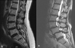
Imaging typically demonstrates a narrow spinal canal, with apparent thickening and buckling of the ligamentum flavum. Degenerative changes of the facet joint are observed as both cartilage loss and the presence of osteophytes with hypertrophy of facet joints. Stenosis in the inferior aspect of the lateral recess (intervertebral foramen) is often seen due to a ventral slip of the superior articular process. The degenerative slip results in reduced foraminal height and more horizontal appearance of the intervertebral foramen at the involved level. The bulging of the degenerated disc into the foramen may further contribute to the foraminal stenosis.23
Natural History
The natural history of degenerative spondylolisthesis (DS) is generally favorable. In a study of 145 non-surgically managed patients with degenerative spondylolisthesis, followed annually for a minimum of 10 years, a progression of vertebral slippage was observed in 34% of the patients.24 However, no correlation between changes in clinical symptoms and progression of spondylolisthesis was observed and low back pain improved following a decrease in the total intervertebral space size. In the study, the development of osteoarthritic spurs, hypertrophy and ossification of the intervertebral ligaments, and facet arthrosis along with loss in disc height was considered to lead to secondary stabilization, thus preventing slip progression. The study also showed that 84 of the 110 patients without neurological deficits at the initial examination remained without neurological deficit at 10-year follow-up. On the other hand, 29 of the 35 patients with neurological symptoms, such as intermittent claudication or vesicorectal symptoms, who refused surgery at baseline were described to have deteriorated neurologically at follow-up.
This study is in agreement with the NASS consensus statement1 presented in 2014 on the natural history of DS, where it was concluded that the majority of patients with symptomatic degenerative lumbar spondylolisthesis and an absence of neurologic deficits will do well with conservative care. Patients who present with sensory changes, muscle weakness or cauda equina syndrome are more likely to develop progressive functional decline without surgical treatment. Slip progression is also less likely to occur when the disc has lost over 80% of its native height and intervertebral osteophytes have formed.
CLASSIFICATION
The grade of spondylolisthesis, can be measured by different methods.25 The first is the method described by Meyerding.26 The anteroposterior (AP) diameter of the superior surface of the lower vertebral body is divided into quarters and a grade of I–IV is assigned to slips of one, two, three or four quarters of the superior vertebra, respectively. Almost all patients with degenerative spondylolisthesis would be graded as grade I or II slips on the Meyerding scale (Fig. 1-1). The second method, described by Taillard,27 expresses the degree of slip as a percentage of the AP diameter of the top of the lower vertebra and is favored by most authors, as it is more accurately reproducible.23 However, measurement of the slip and its apparent progression should be viewed with caution. Studies have shown that the inter- and intra-observer error is up to 15% and that the variation can increase if there is an element of rotation. Therefore, only a progression of greater than 20% slip can be considered a true change using plain radiography.28,29
Besides the radiological classification systems, there is also an etiology-based classification system of all types of spondylolisthesis described by Wiltse et al.30 which includes degenerative spondylolisthesis. However, that classification system made no further attempts to subdivide the degenerative spondylolistheses group. As the pathophysiology and natural history of degenerative spondylolisthesis is much different from the lytic types of spondylolisthesis, it would be desirable to develop a separate system for classification of degenerative spondylolisthesis, which should preferably indicate the severity of the condition as well as some sort of guidance for treatment.
Recently two new classification systems have been proposed for degenerative spondylolisthesis. Kepler et al. have suggested a clinic-radiographic classification, which divides the patients of degenerative spondylolisthesis into 12 subgroups based on the combination of radiographic morphology and leg pain symptoms.31 The classification is as follows:
Morphology subgroups:
Type A: advanced disc space collapse without kyphosis
Type B: disc partially preserved with translation of 5 mm or less
Type C: disc partially preserved with translation of more than 5mm
Type D: kyphotic alignment
Leg pain modifier; No leg pain – 0, Unilateral leg pain – 1, Bilateral Leg pain -2
This classification system can be used for categorization and communication but does not give advice for treatment planning. To address the issues of global lumbar sagittal and coronal plane imbalance, which may have an impact on the treatment decisions for these patients, another classification system has been suggested by the French Society of Spine Surgery.32 This classification divides patients with degenerative spondylolisthesis into 5 subgroups and includes factors of lumbar lordosis (LL), pelvic incidence (PI), pelvic tilt (PT) as well as sagittal vertical axis (SVA):
Type 1: preserved segmental lordosis (> 5◦) and preserved LL (LL > PI-10◦);
Type 2: decreased segmental lordosis (< 5◦) and preserved LL (LL > PI-10◦);
Type 3: decreased LL (LL < PI-10◦);
Type 4: decreased LL (LL < PI < 10◦) with compensation to maintain sagittal balance (PT > 25◦);
Type 5: sagittal unbalance (SVA > 4 cm, with the SVA defined as the distance between the plumb line from the center of the C7 body to the anterior margin of the S1 plate).
For each suggested type the authors present treatment recommendations, however whether using this classification system can lead to a better surgical result remains to be demonstrated.
SUMMARY
In summary, degenerative spondylolisthesis is a condition which is part of the degenerative cascade of the lumbar spine, occurs in middle-aged and older patients and often is diagnosed when presenting with symptoms referring to central and/or lateral spinal stenosis. The natural history of the slippage itself is usually good and the degree of neurological symptoms form the major grounds for decision-making on whether to treat these patients surgically. The classification of degenerative spondylolisthesis has until recently been solely radiological and related to the involved segment(s), however in later years classification systems related to symptoms and other patient factors aim to play a role in the practical care of the patients.
REFERENCES
- NASS Evidence-Based Clinical Guidelines Committee. Diagnosis and treatment of degenerative lumbar spondylolisthesis. 2nd ed. NASS Clinical Guidelines. Burr Ridge, IL; North American Spine Society; 2014.
- Junghanns H. [Spondylolisthesis without gap in the intermediate joint]. Arch Orthop Unfallchir. 1931;29(1):118-127.
- Macnab I. Spondylolisthesis with an intact neural arch; the so-called pseudo-spondylolisthesis. J Bone Joint Surg Br. 1950;32-B(3):325-333.
- Newman PH. Spondylolisthesis, its cause and effect. Ann R Coll Surg Engl. 1955;16(5):305-323.
- Sengupta DK, Herkowitz HN, Degenerative spondylolisthesis: review of current trends and controversies. 2005;30(6 Suppl):S71-S81.
- Kong MH, Morishita Y, He W, et al. Lumbar segmental mobility according to the grade of the disc, the facet joint, the muscle, and the ligament pathology by using kinetic magnetic resonance imaging. 2009;34(23):2537-2544.
- Fitzgerald JA, Newman PH. Degenerative spondylolisthesis. J Bone Joint Surg Br. 1976;58(2):184-192.
- Frymoyer JW. Degenerative spondylolisthesis: diagnosis and treatment. J Am Acad Orthop Surg. 1994;2(1):9-15.
- Iguchi T, Wakami T, Kurihara A, Kasahara K, Yoshiya S, Nishida K. Lumbar multilevel degenerative spondylolisthesis: radiological evaluation and factors related to anterolisthesis and retrolisthesis. J Spinal Disord Tech. 2002;15(2):93-99.
- Aihara T, Takahashi K, Yamagata M, Moriya H, Tamaki T. Biomechanical functions of the iliolumbar ligament in L5 spondylolysis. J Orthop Sci. 2000;5(3):238-242.
- Gao F, Hou D, Zhao B, et al. The pedicle-facet angle and tropism in the sagittal plane in degenerative spondylolisthesis: a computed tomography study using multiplanar reformations techniques. J Spinal Disord Tech. 2012;25(2):E18-E22.
- Rosenberg NJ. Degenerative spondylolisthesis. Predisposing factors. J Bone Joint Surg Am. 1975;57(4):467-474.
- Rothman SL, Glenn WV Jr, Kerber CW. Multiplanar CT in the evaluation of degenerative spondylolisthesis. A review of 150 cases. Comput Radiol. 1985;9(4):223-232.
- Roussouly P, Gollogly S, Berthonnaud E, Dimnet J. Classification of the normal variation in the sagittal alignment of the human lumbar spine and pelvis in the standing position. 2005;30(3):346-353.
- Aono K, Kobayashi T, Jimbo S, Atsuta Y, Matsuno T. Radiographic analysis of newly developed degenerative spondylolisthesis in a mean twelve-year prospective study. 2010;35(8):887-891.
- Barrey C, Jund J, Perrin G, Roussouly P. Spinopelvic alignment of patients with degenerative spondylolisthesis. 2007;61(5):981-986; discussion 986.
- Schuller S, Charles YP, Steib JP. Sagittal spinopelvic alignment and body mass index in patients with degenerative spondylolisthesis. Eur Spine J. 2011;20(5):713-719.
- Funao H, Tsuji T, Hosogane N, et al. Comparative study of spinopelvic sagittal alignment between patients with and without degenerative spondylolisthesis. Eur Spine J. 2012;21(11):2181-2187.
- Kalichman L, Hunter DJ. Diagnosis and conservative management of degenerative lumbar spondylolisthesis. Eur Spine J. 2008;17(3):327-335.
- Jacobsen S, Sonne-Holm S, Rovsing H, Monrad H, Gebuhr P. Degenerative lumbar spondylolisthesis: an epidemiological perspective: the Copenhagen Osteoarthritis Study. 2007;32(1):120-125.
- Sanderson PL, Fraser RD. The influence of pregnancy on the development of degenerative spondylolisthesis. J Bone Joint Surg Br. 1996;78(6):951-954.
- Bird HA, Eastmond CJ, Hudson A, Wright V. Is generalized joint laxity a factor in spondylolisthesis? Scand J Rheumatol. 1980;9(4):203-205.
- Butt S, Saifuddin A. The imaging of lumbar spondylolisthesis. Clin Radiol. 2005;60(5):533-546.
- Matsunaga S, Ijiri K, Hayashi K. Nonsurgically managed patients with degenerative spondylolisthesis: a 10- to 18-year follow-up study. J Neurosurg. 2000;93(2 Suppl):194-198.
- Wiltse LL, Winter RB. Terminology and measurement of spondylolisthesis. J Bone Joint Surg Am. 1983;65(6):768-772.
- Meyerding, HW. Spondylolisthesis: surgical treatment and results. Surg Gynecol Obstet. 1932;54:371-377.
- Taillard W. [Spondylolisthesis in children and adolescents]. Acta Ortho Scand. 1954;24(2):115-144.
- Danielson B, Frennered K, Irstam L. Roentgenologic assessment of spondylolisthesis. I. A study of measurement variations. Acta Radiol. 1988;29(3):345-351.
- Danielson B, Frennered K, Selvik G, Irstam L. Roentgenologic assessment of spondylolisthesis. II. An evaluation of progression. Acta Radiol. 1989;30(1):65-68.
- Wiltse LL, Newman PH, Macnab I. Classification of spondylolisis and spondylolisthesis. Clin Orthop Relat Res. 1976;(117):23-29.
- Kepler CK, Hilibrand AS, Sayadipour A, et al. Clinical and radiographic degenerative spondylolisthesis (CARDS) classification. Spine J. 2015;15(8):1804-1811.
- Gille O, Challier V, Parent H, et al., Degenerative lumbar spondylolisthesis: cohort of 670 patients, and proposal of a new classification. Orthop Traumatol Surg Res. 2014;100(6 Suppl):S311-S315.


