

- See: Radiology of the Pelvis
- Classification:
- elemental frx patterns:
- posterior wall
- posterior column frx
- transverse frx
- anterior column frx
- anterior wall frx
- associated frx patterns:
- T shaped frx
- posterior wall + posterior column frx
- transverse + posterior wall frx
- anterior column or wall + posterior hemitransverse
- posterior column + anterior column frx
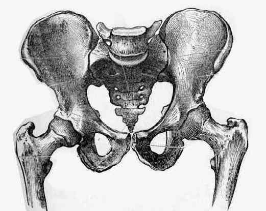
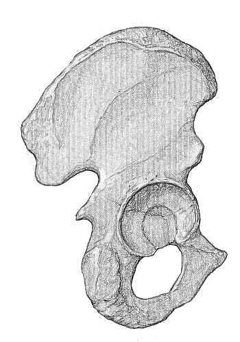
- Radiographs:
- specific views:
- AP view
- Judet View
- landmarks:
- roof arc measurements: determine need for surgery;
- these need to be taken w/ the patient out of traction;
- tear drop
- ilioischial line
- iliopectineal line
- posterior rim
- anterior rim
Predictors for Long-Term Hip Survivorship Following Acetabular Fracture Surgery: Importance of Gap Compared with Step Displacement.
Classification of common acetabular fractures: radiographic and CT appearances
CT of acetabular fractures: postoperative appearances.
Routine pelvic radiography in severe blunt trauma: is it necessary?