

See also:
Discussion
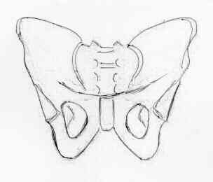
- this view should demonstrate the iliac bone, sacrum, pubis, ischium, femoral heads and necks, and greater or lesser trochanters;
- arcuate, iliopubic, ilioischial, and acetabular teardrop lines, sacral foramina, & SI joint should be scrutinized carefully and identified in a systematic manner;
- patient is supine with the feet slightly (15 deg) internally rotated;
- central beam is directed vertically toward the mid-portion of pelvis;
- for arthritic hips, may get standing films;
- Young and Burgess state that 90 % of all traumatic injuries to the pelvic ring can be diagnosed on AP radiographs alone;
Landmarks include
- iliopectineal line: denotes limit of anterior column;
- ilioischial line: denotes limit of posterior column;
- anterior lip of the acetabulum;
- posterior lip of acetabulum;
- superior wt bearing surface of acetabulum, ending in medial tear drop;
Misc
- in pts w/ severe flexion contractures of hip, AP view frequently appears similar to pelvic inlet view, because pelvis is tilted inferiorly;
- in such cases, better AP radiograph is produced if the hips are slightly flexed to place the pelvis in a more neutral position