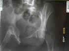
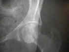
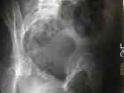
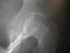
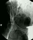
 - See:
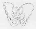
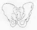
- AP view
- Radiology of the Pelvis:
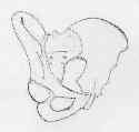
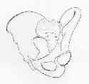
- Internal (Obturator) Oblique View:
- shows iliopectineal line anterior column of pelvis & posterior wall;
- pt is supine w/ involved side of pelvis rotated anteriorly 45 deg & beam directed vertically toward affected hip;
- External (Iliac) Oblique View:
- shows ilioischial line (posterior) column & anterior wall;
- pt is supine w/ uninvolved side of pelvis rotated ant. 45 degrees;
- central beam directed vertically toward the affected hip
- intra-operative flourscopy: it may be difficult to achieve optimal flouroscopy views if the C-arm is placed on the same side as the fracture;
- rotating the injured side to a lower position may improve the view;
Radiographic evaluation of screw position in revision total hip arthroplasty.


