

- See:
- Anterolateral Approach to Forearm
- Forearm Flexors
- Plating Techniques
- Radial Shaft Fractures: Discussion
- Discussion:
- anterior approach of Henry utilizes interval between brachioradialis (radial nerve) & pronator teres (or FCR distally, which are innervated by the median nerve);
- this approach is often used for fractures of the radial shaft, (more often used for fractures in the distal half rather than proximal half);
- disadvantages:
- use of anterior approach (with anterior placement of the plate) may result in block to pronation in proximal third fractures;
- anterior approach requires at least moderate stripping of soft tissues from bone, resulting in delayed return of wrist and hand function;
- Surgical Dissection:
- Incision:
- w/ forearm supinated, begin longitudinal incision at point just lateral & proximal to biceps tendon (at the flexor crease of the elbow) extend it distally in forearm along medial border of brachioradialis towards the radial styloid;
- expose the biceps tendon by incising deep fascia on its lateral side;
- incise biciptial bursa, which lies in angle between lateral margin of biceps tendon & the radius;
- then divide deep fascia of forearm in line w/ skin incsion, taking care to protect the radial vessels;
- fascia is incised between brachioradialis & FCR;
- preserve lateral cutaneous nerveantebrachial lateral nerve which lies subcutaneously;
- superficial branch of the radial nerve lies along undersuface of the brachioradialis, which is protected by lateral retraction of the BR;
- proximal exposure:
- Henry approach can also be modified to allow exposure to the anterior elbow and proximal radius;
- if more proximal exposure is required, then dissect between the brachioradialis and the brachialis (radial nerve will lie between these muscles);
- the exposure can be further extended w/ the anterior approach to the humerus;
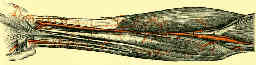
- Isolation of the Radial Artery:
- the radial artery is best identified distally and followed proximally;
- once the distal artery has been found, the brachioradialis & superficial radial nerve are retracted radially revealing the proximal portion of the radial artery;
- radial artery lies beneath brachioradialis in middle part of forearm, and lies close to medial edge of wound;
- artery may have to be mobilized & retracted medially to achieve satisfactory exposure of deeper muscular layer;
- it runs w/ two venae comitantes, which remain prominent if limb is not exsanguated before the tournequet is applied;
- because the radial artery is vulnerable during mobilization of brachioradialis, its branches to the brachioradialis must be ligated (bipolar cautery);
- proximal mobilization of the brachioradialis requires ligation of the recurrent radial artery;
- Deep Dissection:
- BR is retracted laterally and the pronator teres is retracted medially;
- flex elbow to right angle to allow more complete retraction of brachioradialis & radial carpal extensor to expose supinator;
- brachioradialis is supplied by several branches of radial artery which are ligated;
- superficial branch of the radial nerve lies along undersuface of the brachioradialis, which is protected by lateral retraction of the BR;
- more distally, the dissection procedes between the brachioradialis and the FCR which is also retracted medially (along w/ the pronator);
- Dissection of the Forearm Muscles Off the Radius:
- supinator:
- proximally, fibers of supinator are identified as are fibers of pronator teres which will be seen more distally passing over supinator in opposite direction;
- proximally, supinator is incised at its insertion on radius, and subperiosteally stripped from radius;
- supination of the forearm displaces the PIN laterally, away from operative field;
- supinator insertion should be exposed in full supination & detached from radius as close as possible to the bone;
- through an incision starting at the flexor crease of elbow & following medial border of brachioradialis belly distally toward the lower forearm;
- supinator is then is reflected laterally together w/ deep branch of radial nerve (PIN) w/ in its substance (lateral retraction protects the PIN);
- for more proximal exposure, begin dissection of the supinator origin just lateral to the biceps tendon;
- isolate & ligate leash of Henry & subperiosteally strip supinator from its insertion;
- FDS:
- the FDS insertion begins just distal to the bicipital tuberosity and is ulnar to the supinator;
- pronator teres:
- in middle third, insertion of pronator teres muscle is preserved if possible;
- if dissection is required, pronate the arm to better expose its insertion;
- FPL & PQ
Vulnerability of the posterior interosseous nerve during proximal radius exposures.
Anatomic considerations for the anterior exposure of the proximal portion of the radius.