

- Discussion:
- main function of the tibialis posterior is to invert the subtalar joint, which helps to stabilize the transverse tarsal joint during
rheumatoid hindfoot;
- pathophysiology:
- degenerative tears usually occur distal to the medial malleolus, in a region which coincides with relative hypovascularity and
sharp turn of the tendon from a verticle to a horizontal position;
- as the tendon makes a sharp turn under the medial malleolus, there may be fibrous metaplasia, leading to rupture;
- in the study by Mosier SM, et al (1998), gross and histological exams were carried out on 15 normal cadvaers and 15
surgical patients w/ posterior tibial tendon insufficiency (but no rupture);
- these authors noted that 12/15 cadavers had normal tendon appearance and histology, where as the surgical specimens
demonstrated a degenerative tendinosis w/ increased mucin content, fibroblast hypercellularity, chondroid
metaplasia, and neovascularization
(the abnormal tendon segments were located between the medial malleolus and the navicular tuberosity;;
- gross examination of the surgical specimens showed incomplete splitting on the deep surface;
- following rupture, talonavicular joint and subtalar joints collaspes, & hindfoot drifts into valgus, causing mid foot pronation
& forefoot abduction;
- in addition, there is often injury or attenuation of the spring ligament;
- in the rheumatoid, rupture of the tibialis posterior leads to a collapsed pronated foot;
- ref:
Pathology of the posterior tibial tendon in posterior tibial tendon insufficiency..
- clinical manifestations:
- early in this condition there is painful swelling along posteromedial border of ankle, fatigue, & aching along medial
longitudinal arch of foot;
- well into disease process, patients may note lateral sided pain as well as pain in sinus tarsi impingement between lateral side
of foot and fibula;
- in advanced stages, pain is present laterally, w/ an abutment between the calcaneus and the fibula;
- peroneus brevis, continues to function & pulls foot into a valgus configuration;
- in this case, flat foot may result from the peroneus brevis muscle, which is a natural antagonist to the tibialis posterior;
- rupture classification:
- stage I
- stage II
- stage III
- stage IV:
- Update on stage IV acquired adult flatfoot disorder: when the deltoid ligament becomes dysfunctional.
- diff dx:
- attenuation of the spring ligament;
- rheumatoid foot:
- look for involvement in the hindfoot and talonavicular joints;
- synovitis and joint inflammation lead to weakening of these joints which results in hindfoot valgus deformity which
resembles rupture of TP;
- tarsometatarsal degenerative arthitis
- relaxed pes planus
- old lisfranc fracture dislocation
- neuroarthropathic (charcot) involvement of the midfoot or hindfoot
- posteromedial talar osteochondral lesion;
- associated conditions:
- most patients will not have an associated condition;
- rheumatoid arthitis;
- seronegative arthritis;
- Radiographs:
- wt bearing lateral;
- talus moves into flexion when viewed laterally; 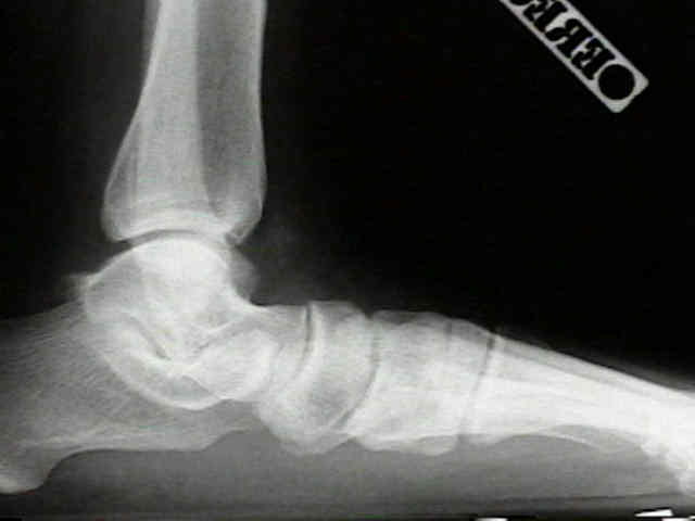
- talus will appear plantar flexed and there will be an increased angle between
longitudinal axis of the talus and calcaneus;
- decrease in the talometatarsal angle (normally 0 to 10 deg);
- wt bearing AP view;
- talar head uncoverage and increase in angle between longitudinal axis of talus & calcaneus
- the displacement of forefoot into abduction w/ calcaneus is assessed;
- lateral subluxation of the navicular on the talus correlates w/ the amount of deformity;
- MRI:
- ref: Foot Ankle. 1992 May;13(4):208-14.
Clinical significance of magnetic resonance imaging in preoperative planning for reconstruction of posterior tibial tendon ruptures..
- Non Operative Rx:
- use of arches supports, and heel cups, is usually unsuccessful in providing relief from symptoms of foot strain in these pts;
- posterior tibial tendonitis:
- objective is to reduce excessive midfoot motion;
- consider total contact orthosis supporting longitudinal arch;
- medial heel wedge;
- references:
- Nonoperative treatment of patients with posterior tibial tendinitis. Lin, et al. Foot and Ankle Clin. 1996;1:261-277.
- Nonoperative treatment of posterior tibial tendon pathology. Sferra, et al. Foot and Ankle Clin. 1997;2:261-273.
- Nonoperative management of posterior tibial tendon dysfunction.
- Surgical Treatment:
- tenosynovectomy:
- indicated early in disease process (stage I - prior to rupture) after the patient has failed a trial of immobilization;
- tenosynovectomy is performed w/ care to preserve a portion of the flexor retinaculum (to prevent subluxation of the tendon);
- tendon sheath is opened and the tendon is inspected;
- pathologic tissue is removed;
- flexor retinaculum is not closed (assumming a significant portion remains intact to prevent subluxation);
- FDL transfer and osteotomy:
- often combined w/ repair or reconstruction of the sping ligament;
- achilles tendon lengthening may also be required;
- medial displacement calcaneal osteotomy;
- shifts the achilles tendon medial to the axis of the subtalar joint, which helps support the tendon transfer medially;
- this procedure may reduce the lever arm acting across the subtalar joint;
- this procedure may not be of much benefit in patients w/ severe forefoot abduciton;
- lateral column lengthening;
- references:
- Surgical treatment of stage II posterior tibialis tendon dysfunction: ten-year clinical and radiographic results.
- Radiographic Outcomes Following Lateral Column Lengthening With a Porous Titanium Wedge
- arthrodesis:
- talonavicular joint arthrodesis:
- if site of maximal deformity is at talonavicular joint, then an isolated talonavicular or talonavicular & calcaneocuboid may
be performed;
- sub-talar arthrodesis:
- w/ more significant hindfoot valgus, subtalar arthrodesis may be indicated.
- note that complete correction of the hindfoot deformity may cause a relative supination deformity of the
forefoot (which in turn, decreases the relative amount of wt bearing of the first metatarsal);
- triple arthrodesis:
- indicated only w/ severe midfoot collapse deformities;
- achilles tendon lengthening will often be required for equinus deformity;
Rupture of the posterior tibial tendon associated with closed ankle fracture.
Acquired adult flat foot secondary to posterior tibial-tendon pathology.
Rupture of the tibialis posterior tendon.
Rupture of the posterior tibial tendon causing flat foot. Surgical treatment.
Tibialis posterior tendon dysfunction.
Posterior tibial tendon dysfunction: its association with seronegative inflammatory disease.
Pathology of the posterior tibial tendon in posterior tibial tendon insufficiency.