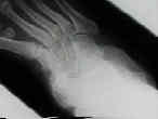
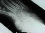
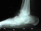
(see also: Blair Fusion; Charcot Marie Tooth ; Equinovalgus; Grice Arthrodesis; Kocher approach; Ollier incision; Subtalar Fusion)
Discussion
- results in fusion of subtalar, calcaneocuboid, and talonavicular joints;
- most effective procedure for fixed hindfoot and forefoot deformities;
- triple arthrodesis tend to have a high (50%) failure rate in children under 10 years of age;
- triple arthrodesis in polio (see polio):
- in polio pt, the goals of triple arthrodesis:
- stable static realignment of foot;
- removal of deforming forces
- arrest of progression of deformity
- elminination of pain and decrease limp;
- normal appearing foot;
Stiffness following islolated arthrodesis
- subtalar arthrodesis limits talonavicular motion to about 26%, and limits calcaneocuboid motion to about 56%;
- talonavicular fusion will limit subtalar motion to about 9% so that there will only be about 2 deg of motion;
- calcaneocuboid fusion decreases talonavicular motion down to 67% and subtalar motion down to 92%;
- reference: Selective tarsal arthrodesis: An in vitro analysis of the effect on foot motion.
Outcomes
- Saltzman CL, et al (1999): 67 feet in 57 patients who underwent triple arthrodesis were evaluated at an average of 25 and 44 years following surgery;
- indications for surgery: polio (55%), CMT (9%), spinal cord abnormalities (6%), CP (4%), Guillain-Barré syndrome (1%);
- pseudarthrosis occurred in thirteen feet
- 55 % of feet were painful at 44 years and all showed some degenerative changes;
- despite the foot pain and radiographic signs of degenerative changes, 95% of patients were satisfied with results of the procedure;
- the authors note that the indications for triple arthrodesis have change during the past few decades (less often for neuromuscular disorders and more often for severe degenerative arthritis or severe flat foot), and note that the prevalence of pseudoarthrosis is less frequent;
- Pell RF, et al. (2000), the authors followed 183 triple arthrodeses procedure over an average of 5.7 years;
- authors noted high correlation between patient satisfaction and post-operative foot alignment;
- 91% of patients indicated that they would have the procedure again, under similar circumstances;
- postoperative ankle arthritis (clinical and radiographic) was common but did not seem to affect patient satisfaction;
- feet w/ post-traumatic changes (25) required the least correction where as the 22 feet w/ RA and the 70 feet w/ PT rupture needed the most correction;
- average time to fusion was 10.7 weeks;
- Triple arthrodesis: twenty-five and forty-four-year average follow-up of the same patients.
- Clinical Outcome After Primary Triple Arthrodesis
Contra-indications
- contra-indicated in young children (less than 10-12 yrs) because the procedure limits foot growth;
Preoperative Planning
- Surgical Instruments:
currettes, lamina spreader w/ and w/o teeth, osteotomes, peri-osteal elevators, spade retractors, homan retractors, and curved creigo elevators; - Operative Technique:
- positioning:
- supine, w/ bumb under hip;
- prep for bone graft;
- soft tissue considerations:
- consider need for peroneal tendon lengthening and achilles tendon lengthening (equinus contracture);
- posterior ankle capsular release:
- posteromedial incision parallel to the Achilles tendon, retract FHL medially, and capsule is incised;
- varus deformity:
- release the medial talonavicular capsule;
- medial subtalar capsule requires release;
- navicular and calcaneus are then brought back around the talus;
- incision:


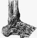


- EDB is freed from its insertion & reflected distally, w/ care to preserve the nerve supply;
- adjacent to lateral border of EDB is peroneal tendon sheath;
- medial extent of EDB is adjacent to peroneus tertius tendon;
- proximal extent of EDB is noted as it disappears into sinus tarsi, beneath the fat pad and the inferior extensor retinaculum;
- outline of EDB is completed allowing creation of a distally based EDB flap;
- in medial & dorsal aspects of EDB, keep dissection subperiosteal to avoid neurovascular damage;
- NV bundle enters approx 1.5 cm medial & distal to anterior process of the calcaneus;
- retract peroneal tendons out of their tendon sheath plantarly & laterally;
- continue dissection to cuboid and distal calcaneus thru deep portion of peroneus sheath;
- EDB belly is delivered out of Sinus Tarsi by sub-perosteal dissection;
- remove the fibro-fatty contents of the sinus taris w/ a rounguer;
- identify, posterior facet of the subtalar joint;
- identify the calcaneocuboid joint which is found just distal to the anterior beak of the calcaneous;
- avoid entering into the joint;
- the talo-navicular joint lies superior and medial to calcaneocuboid joint;
- identify neck of the talus, and elevate the extensor tendons off the neck;
- insert a spade over the neck / under the extensor tendons which helps retract the N/V bundle;
- joint capsule of lateral talar neck is dissected in plantar to dorsal direction subperiosteally then moving distally to area of navicular;
- positioning:
Resection of Subtalar Joint
- bear in mind that resection of this joint will establish the position of hindfoot varus-valgus position;
- interosseous ligament was divided which allowed access to each facet of the subtalar joint
- bifurcate ligament was released to provide full correction of the abduction deformity of the midfoot
- insert a Creigo curved elevator deep to the peroneal tendon and around the calcaneus at the level of the sub talar joint;
- consider removing the lateral talar process to better expose the posterior facet;
- joint capsule of talocalcaneal joint is incised & laminar spreader is inserted into sinus tarsi to expose entire subtalar articulation;
- anterior facet of the subtalar joint is found by moving inferior to the talar head;
- anterior facet is a relatively medial structure and may be in direct communication with the talonavicular joint;
- excise articular cartilage & subchondral bone of subtalar joint:
- care should be taken to insure excision of articular catilage from anterior, middle, and posterior facet;
- all of the non articular surfaces are decorticated;
- avoid excessive bone resection since this will decrease the subtalar joint height and will disrupt articular relationship of the talo-naviulcar joint;
- when debriding posteromedial aspect of the joint, use currette instead of the hall drill to avoid impailing the NV bundle;
- flexing great toe, will result in movement of FHL tendon is helpful in localizing the exact location of N/V bundle since it lies anteromedial to FHL at level of subtalar joint;
Resection of Calcaneocuboid Joint
- bifurcate ligament is released to provide full correction of the abduction deformity of the midfoot
- lateral column lengthening:
- when indicated, distraction arthrodesis of the calcaneocuboid joint is performed with interposition bone graft;
Resection of Talo-navicular Joint
- bifurcate ligament is released to provide full correction of the abduction deformity of the midfoot
- resection of this joint will partially establish supination / pronation of the forefoot;
- insert spade retractor over the talar neck to protect NV bundle;
- w/ planovalgus deformity, manually reduce head of talus into the lateral wound;
- if reduction cannot be achieved or exposure is inadequate consider a circumferential release of talonavicular joint;
- note that talar head is made of softer bone than navicular, which creates path of lesser resistance for bur;
- note that medial aspects of talonaviular joint curves well away from wound, making visualization difficult;
Medial incision (optional)
- incision is made dorsomedially, beginning at tip of medial malleolus and in line w/ medial border of foot, extending 6-7 cm toward great toe longitudinally & ending between anterior & posterior tibial tendons;
- saphenous vien is retracted dorsally;
- subperiosteal dissection is carried out plantarly and dorsally along navicular;
- dorsal dissection is limited inorder to preserve blood supply to talus;
Cautions
- talonavicular joint is a round joint that follows the contour of the talar head;
- only the medial two thirds of the joint can be reached from the medial incision (remainder is exposed via the lateral approach);
- poor exposure (not exposing the entire talonavicular joint) may explain high nonunion rate for talonavicular fusion;
- reach the lateral side of the talonavicular joint by following the lateral talar neck distally;
IntraOp Position of the Foot
subtalar joint reduction:
- when reducing subtalar joint in the case of a planovalgus foot, it is not simply varus ward correction but also internal rotation of calcaneus underneath the talus;
- in the report by Maenpaa H, et al, 307 triple arthrodeses were done on 282 patients with rheumatic diseases between 1995 and 1999;
- solid and painless fusion was achieved in 261 patients (93%, 286 arthrodeses);
- 21 arthrodeses (in 21 patients) that failed were analyzed;
- 14 (66%) malunions, 6 (29%) nonunions, and one (5%) painful foot without malunion or nonunion were found;
- of the failed procedures, valgus alignment was present in 13 feet and varus alignment was present in eight feet;
- most common cause of failure was a misjudgment in the surgical technique, which occurred in 12 of 21 (57%) patients based on inadequate correction and repositioning of hindfoot deformity;
- in four (19%) patients, additional ankle destruction and instability was overlooked as a cause of malalignment;
- revision triple arthrodesis was successful in 18 of 21 (86%) patients;
- ref: What Went Wrong in Triple Arthrodesis? An Analysis of failures in 21 Patients
planovalgus deformity
- consider lateral column lengthening vs. medial column shortening;
- typically when the foot is reduced, a gap will be present at the calcaneocuboid interval, and will require bone graft;
- in a sense, it is a lateral column lengthening by necessity;
equinovarus deformity
- consider pining the talonavicular joint w/ the talus in flexion so that the ankle will not impinge in dorsiflexion;
- w/ fixed midfoot supination, the navicular may have to be depressed in relation to the talus, to help spin the medial side of the foot down into a plantigrade position;
- in this case, the superior aspect of the talus will be uncovered;
- consider removing the superior 1/3 of the talus, not only to to prevent ankle impingement in dorsiflexion, but also to obtain more bone graft;
Fixation 
subtalar joint
- correction of hindfoot valgus is achieved at subtalar joint;
- anterior to posterior screw insertion:
- insert pin medial to anterior tibial tendon from talar neck into calcaneus;
- if fixation screw is inserted w/ ankle in plantar flexion and is located too close to anterior margin of tibia, it may block ankle dorsiflexion or alternatively may irritate the ankle joint;
- usual length of screw is 65 mm long;
- visualized pin as it goes thru sinus tarsi region into calcaneus;
- bulge of the pin under the skin of the heel region is then felt and the pin is retracted back about 2 cm;
- advantages: there is better fixation w/ this method (compared to the posterior to anterior insertion), since long threaded lag screws can be used;
- disadvantages: if the screw is position too close to the talar head, then painful ankle impingement may occur when the ankle is placed in maximum dorsiflexion;
- posterior to anterior screw insertion:
- alternatively, the Steinman pin or 7.0 cannulated screw is inserted from the heel pad, across subtalar joint, into distal tibia;
- it is best to insert the screw just superior to the wt bearing portion of the heel;
- advantages: this method will avoid impingement of the screw head when the ankle is placed in dorsiflexion;
- disadvantages: the lag screw is placed from the larger fragment into the smaller fragment and therefore lag screws w/ short threads must be used;
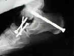
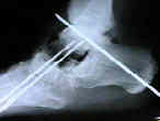
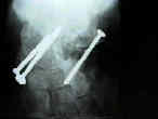
- alternatively, the Steinman pin or 7.0 cannulated screw is inserted from the heel pad, across subtalar joint, into distal tibia;
Talo-navicular joint
- correction of forefoot rotation;
- insert pin from naviculo-cuneiform joint toward center of head of talus;
- w/ optimal positioning, the talar neck, navicular, and 1st metatarsal will be co-linear;
- fixation of the talonavicular joint was performed first using two screws: (screws are inserted from navicular into the talus);
- one lag screw from the tubercle of the navicular into the talus
- starting point needs to be distal on the navicular (create a notch can be made in the medial cuneiform) to get exposure;
- second cortical screw from the neck of the talus into the lateral part of the navicular
- through a small incision dorsolaterally
- one lag screw from the tubercle of the navicular into the talus
Calcaneocuboid Joint
- lateral column lengthening:
- typically is the last joint too be fixed;
- correction of forefoot abduction / adduction;
- fixation can be achieved w/ two crossed 4.5 mm cannulated screws;
- usually fixation must be directed from the anterior process of the calcaneus to the cuboid;
Final Assessment and Application of Bone Graft
- after fixation is completed, foot and heel is assessed for alignment;
- if needed calcaneal osteotomy is performed for residual calcaneal varus or valgus angulation after performing the arthrodesis;
- bone graft is applied;
Post-Operative Care
Complications
- pseudoarthrosis of talonavicular joint;
- may be due to the fact that the talonavicular joint has more motion than either the subtalar joint or the calcaneocuboid joint;
- forefoot deformity (supination / pronation) which requires first metatarsal - cuneiform osteotomy and fusion
References
- Hoke triple arthrodesis.
- Triple arthrodesis in older adults. Results after long-term follow-up.
- Long-term results of triple arthrodesis in Charcot-Marie-Tooth disease.
- Triple arthrodesis in rheumatoid arthritis.
- Triple arthrodesis using internal fixation in treatment of adult foot disorders.
- Triple arthrodesis in adults.
- Triple arthrodesis. A critical long-term review.
- Clinical Outcome After Primary Triple Arthrodesis
- Growth rates in skeletally immature feet after triple arthrodesis.
- Single medial approach to modified double arthrodesis in rigid flatfoot with lateral deficient skin.


