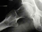
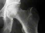
 - AP view:
 - patient is supine with the foot internally rotated 15 deg to obtain best views of the femoral neck;
- central beam is directed toward the femoral head;
- X-ray tube should be positioned 100 cm from focal plane of film cassette to yield an image at 20%
magnification, corresponding to the magnification incorporated in the templates;
- tape measure will allow accurate assessment of radiographic magnification;
- Lateral View:
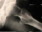
- surgical lateral view:
- this view should be obtained on all patients suspected of having a hip fracture or dislocation;
- do not order a frog leg lateral in any patient suspected of having a hip fracture or dislocation)
- patient is supine; the opposite hip is flexed and abducted;
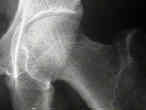
- cassette is placed against the lateral aspect of the affected hip;
- central beam is directed horizontally toward the groin with about 20 degree of cephalic tilt;
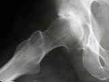
- frogleg lateral view:
- do not order a frog leg lateral in any patient suspected of having hip fracture or dislocation);
- patient is supine w/ knees flexed, soles of feet together, and the thighs maximally abducted;
- central beam is directed vertically or with a 10 to 15 deg cephalic tilt to a point slightly above pubic symphysis;
- Radiographic Evaluation for Hip Arthroplasty:
- preop x-rays for THR:
- post op x-rays for THR:
- Radiographic Evaluation of Femoral Neck Fractures:
- Radiographic Evaluation for Acetabular Fractures
- judet views
- roof arc measurements:
- Radiographic Evaluation for Pelvic Frx
The lateral trochlear sign. Femoral trochlear dysplasia as seen on a lateral view roentgenograph.
A Systematic Approach to the Plain Radiographic Evaluation of the Young Adult Hip


