

- See:
- Bone Patella Bone Allografts
- Non Bone Patella Bone Reconstruction
- Discussion:
- Anatomy of ACL;
- ACL Biomechanics;
- Graft Tunnel Sites / Isometry:
- Graft Biomechical Characteristics:
- according to Noyes et. al., strength of 14 mm-wide graft of patellar tendon is approximately 175% the strength of normal ACL;
- in most cases a 9 or 10 mm wide graft is used, and thus the strength is less (112% of normal);
- semitendinosus graft is approx 75%, gracilis tendon is approx 25%, and 20 mm-wide iliotibial band is approx 20% strength of
normal ACL;
- when each of these grafts is doubled over, however, the strength may be increased to 250% of normal;
- while ultimate tensile strength is important, of greater significance may be graft stiffness;
- PreOp Evaluation:
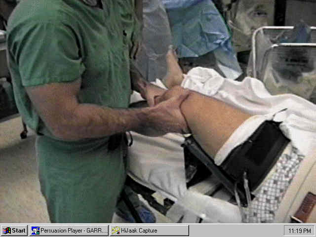
- Positioning and Setup:
- Arthroscopic Technique:
- portals: (see knee arthroscopy)
- medial and lateral portals need to be placed on either side of the patellar tendon;
- bleeding in ACL reconstruction procedures is more common than w/ standard arthroscopy since there is often an angry hemorrhagic
syovitis around the intercondylar notch and because soft tissue debridement along the lateral wall stirs up bleeding;
- joint and the subcutaneous tissues over the portal sites are infiltrated w/ 30 ml of 0.25% bupivacaine w/ epinephrine solution (1:200,000);
- inaddition to a dedicated inflow cannula, consider applying a second inflow cannula to the arthroscope;
- hence if knee flexion decreases flow from the suprapatellar pouch, there will still be flow arising from the arthroscope;
- prior to debriding the intercondylar soft tissues, ensure that the inflow is optimal;
- if the superior fat pad is hypertrophic, then it may have to be debrided inorder to allow passage of fluid from
the inflow cannula down into the notch;
- it is also helpful to place the tip of the inflow cannula directly over the trochlea;
- have intra-articular cautery available and ready to deal with bleeding soft tissues;
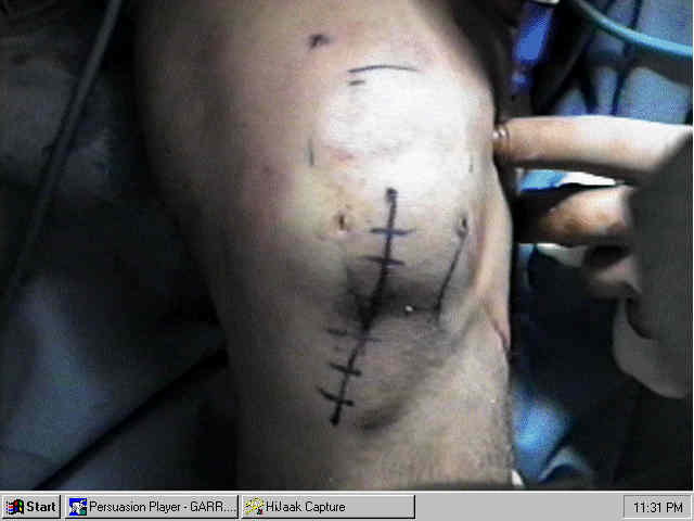
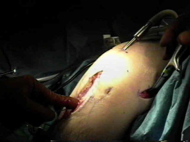
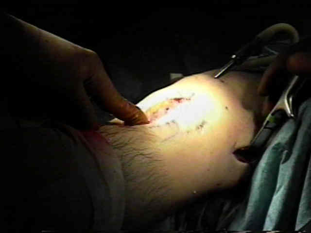
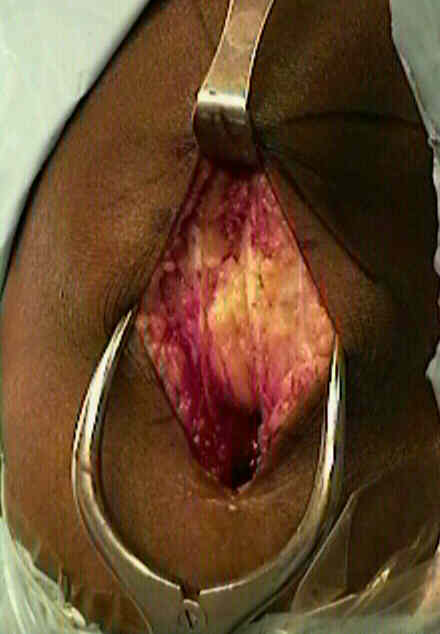
- Patellar Tendon Harvest and Preparation:
- Patellar Tendon Harvest:
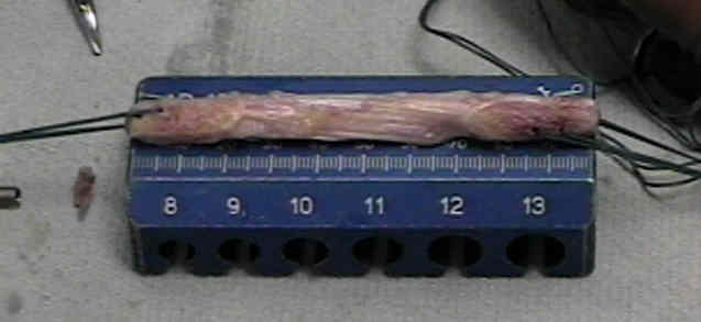
- Patellar Tendon Graft Preparation:
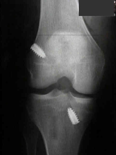
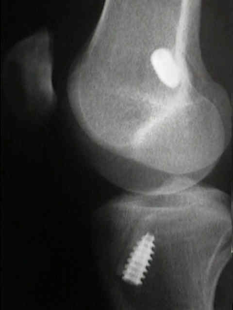
- Tibial Tunnel:
- Femoral Tunnel:
- NotchPlasty:
- intercondylar notch area is then cleared using an osteotome and/or a 5.5 mm shaver;
- anterolateral notchplasty should remove approximately 3-5 mm;
- One Incision Technique:
- main disadvantage is inability to restore the anatomy of the femoral ACL attachment;
- Two Incision Technique:
- probably the gold standard of ACL reconstruction, because it most closely restores the anatomy of the femoral ACL attachment
Graft Passage and Fixation:
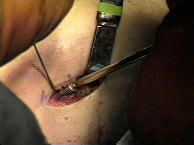
- Graft Passage:
- prior to graft passage ensure that an adequate notchplasty has been performed by passing an appropriately sized smooth metal rod
through the tibial tunnel;
- w/ the knee in extension, the rod should easily pass into the notch;
- Graft Fixation:
- prior to distal (tibial) graft fixation, determine whether the graft changes position as the knee is taken from 90 deg flexion to full
extension;
- then cycle the knee 15-20 times for stress relaxation;
- normal ACL has approximately 3 mm of excursion from 30 deg of flexion to 0 deg extension; (Garrett and Cavallo, unpublished data);
- in a typical ACL reconstruction there will be 2-3 mm of excursion from 30 to 0 deg of extension;
- hence graft should be fixed w/ the knee in 20-30 of flexion, so that the physiologic tension can be maintained;
- ref: Effects of Twisting of the Graft in Anterior Cruciate Ligament Reconstruction.
 - Wound Closure:
- attempt to collect all bone chips created from reaming the femoral and tibial tunnels;
- these bone chips may be applied to the patellar harvest site to hasten defect healing;
- it is noteworthy that in many cases the patellar bone defect will remain unfilled for several months postoperatively,
and this stress riser may account for anecdotal accounts of late patellar fracture following ACL reconstruction;
- some surgeons elect to repair the patellar tendon w/ 0 Vicryl simple or horizontal matress sutures (avoid figure of 8
because this may produce patellar baja);
- paratenon is closed using 2-0 Vicryl running suture.
- some surgeons do not repair the patellar tendon at all;
- in either case, care should be taken to repair the paratenon;
- references:
- Infrapatellar contracture syndrome: diagnosis, treatment, and long term follow up.
- Anterior cruciate ligament patellar tendon reconstruction: it is probably better to leave the tendon defect open!
- Post Op Care of ACL Reconstructions
Intra-articular cruciate reconstruction. II: Replacement with vascularized patellar tendon.
Fractures associated with patellar ligament grafts in cruciate ligament surgery.
Vascularized patella tendon anterior cruciate ligament reconstruction.
Use of Allografts after Failed Treatment of Rupture of the Anterior Cruciate Ligament.
Functional anatomy of the anterior cruciate ligament and a rationale for reconstruction.