

- Discussion:
- there is inherent graft weakness during period after implantation when allograft& autograft tissues undergo necrosis,
revascularization, and cellular proliferation;
- weakest link initially is graft fixation site;
- it is important to achieve strong fixation initially;
- bone plugs with interference screws seem to provide greatest fixation strength;
- as healing occurs, strength at fixation sites > graft strength;
- normal ACL has approximately 3 mm of excursion from 30 deg of flexion to 0 deg extension; (Garrett and Cavallo, unpublished data);
- in a typical ACL reconstruction there will be 2-3 mm of excursion from 30 to 0 deg of extension;
- hence graft should be fixed w/ the knee in 20-30 of flexion, so that the physiologic tension can be maintained;
- note: when a graft appears to have no excursion (w/ flexion and excursion) always consider whether
the graft is binding up within the tibial tunnel;
- bioabsorbable screws:
- motivation for bioabsorbable screws include the following:
- no need for hardware removal;
- no metal artifact interferance with a future MRI;
- possible reduced occurance of suture laceration by the screw threads;
- polylactide screws may be more biocompatible than polyglylactide screws;
- in the study by Tuompo et al (1996), there was better fixation strength with a standarad metal screw,than seen with the bioabsorable
screws, however, the absorbable screws had a pull out strength of 1300 to 1400 N, which was considered accetable;
- in the biomechanical study by Johnson et al. (1996), the load failure for a metal screw was 436 N vs 565 N for a bioabsorbable
screw;
- refs:
- Strength of the fixation of patellar tendon bone grafts using a totally absorbable self reinforced Poly-L-Lactide expansion plug and screw. An experimental study in a Bovine Cadaver.
- Metal and biodegradable interference screws: comparison of failure strength
- Technique:
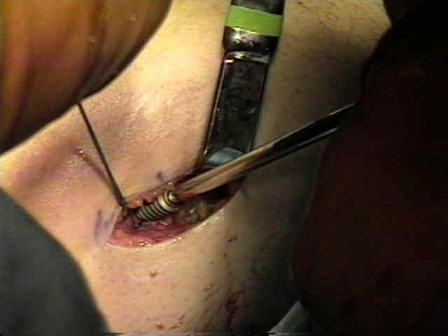
- ensure that the cortical side of the proximal bone plug faces posteriorly;
- look for the blue mark at the tendon-bone edge of the plug to ensure that the entire plug resides with in the femoral tunnel;
- femoral plug fixation:
- Arthrex Tunnel Notcher can be used to create a notch in the anterior portion of the tunnel to allow insertion of the guide wire parallel
to the bone plug graft;
- both the Notcher and the guide wire can be inserted thru the antero-medial portal or may be inserted through a separate portal
inserted the the patellar tendon harvest defect (thru the fat pad);
- the guide wire is inserted parallel to the bone plug, to a depth of at least 25 mm;
- if the interference screw diverges as little as 15 deg from the graft direction, the strength of fixation will be compromised;
- choose a interference screw that is 2 mm smaller than the tunnel diameter, which is slightly smaller than the length of the plug;
- firm tension is applied the the proximal bone plug sutures;
- the interference scew can be inserted over a plastic sheath which will facilitate controlled insertion of the screw over the wire;
- careful arthroscopic visualization is required during screw insertion;
- avoid screw divergence:
- note that use of the endoscopic transtibial technique risks screw insertion at a divergent angle;
- divergence of more than 15 deg risks significant weakening of the bone plug pull out strength;
- references:
- Femoral interference screw placement through the tibial tunnel: a radiographic evaluation of interference screw divergence angles after endoscopic anterior cruciate ligament reconstruction.
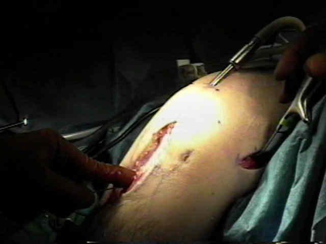
- pre-tension and test the graft:
- firm tension is applied to the bone plug sutures and the knee is taken thru 15-20 flexion-extension cycles (to allow for stress
relaxation);
- subsequently, attempt to judge the graft excursion during flexion-extension;
- ACL graft should pull up into the tibial tunnel by about 2mm w/ extension when fixed on the femoral side;
- this represents the closest reconstitution of the ACL's "physiometry" (see: isometry);
- a graft that tightens (pulls up into the tibial tunnel) with flexion will have a much higher likelyhood of failure and usually indicates a
femoral tunnel too far anterior in the notch;
- generally, the graft is placed in maximum tension at 20-30 deg flexion, while a posterior drawer stress is placed on the tibia;
- tibial interference screw size:
- generally, the tibial tunnel interference screw should be 1 mm less than the size of the tunnel (a larger screw is needed because there
is often slight widening of the tibial tunnel as instruments are passed thru it in order to complete drilling of the femoral tunnel);
- tibial bone plug position:
- the general goal is to have the cortical portion of the graft positioned anteriorly and the interference screw inserted posteriorly in the
tunnel;
- if it appears that there is graft impingement, then consider placing the interference screw anteriorly, in order to move the graft
slightly posteriorly;
- graft rotation:
- the tibial bone plug can be rotated 90 deg internally, to reproduce femoral anatomy;
- this is especially useful if the graft is slightly long;
- this also makes it less likely that the screw threads will cut thru the graft sutures during the interference screw fixation;
- references:
- The effects of graft rotation on attachment site separation distances in anterior cruciate ligament reconstruction.
- tibial side fixation:
- if the interference screw diverges as little as 15 deg from the graft direction, the strength of fixation will be comprimised;
- in the report by Höher J et al., 10 cadaveric knees were tested at full extension, 15°, 30°, and 90° of flexion under a 134-N anterior tibial load;
- in each knee, the kinematics as well as in situ force in the graft were compared when the graft was fixed with the tibia in four different positions:
- full knee extension while the surgeon applied a posterior tibial load (Position 1);
- 30° of flexion with the tibia at the neutral position of the intact knee (Position 2);
- 30° of flexion with a 67-N posterior tibial load (Position 3);
- 30° of flexion with a 134-N posterior tibial load (Position 4);
- for positions 1 and 2, the anterior tibial translation and the in situ forces were up to 60% greater and 36% smaller, respectively,
than that of the intact knee;
- for Position 3, knee kinematics and in situ forces were closest to those observed in the intact knee;
- for Position 4, anterior tibial translation was significantly decreased by up to 2 mm and the in situ force increased up to 31 N;
- these results suggest that the position of the tibia during graft fixation is an important consideration for the biomechanical
performance of an anterior cruciate ligament-reconstructed knee.
- ref: The Position of the Tibia during Graft Fixation Affects Knee Kinematics and Graft Forces for Anterior Cruciate Ligament Reconstruction
- graft should be fixed w/ the knee in 20-30 of flexion, so that the physiologic tension can be maintained;
- a posterior drawer stress is not esssential, but firm tension needs to be applied to the graft sutures;
- the main pitfall to consider is that the interference screw may push the graft proximally, which will cause loss of graft tension;
- progress with screw insertion can be check by placing the arthroscope in the tibial tunnel;
- screw position:
- the interference screw may be inserted slightly medially or laterally along the posterior surface of the tunnel in order to avoid
cutting the bone plug sutures;
- if the interference screw is inserted in the anterior poriton of the tunnel, it may fracture through the anterior cortical surface;
- tibial washer fixation:
- in the report by Post WR, et al, the authors sought to define the posterior neurovasculature structures at risk when drilling for
bicortical tibial screw fixation during ACL reconstruction;
- the authors placed the tibial tunnel arthroscopically in 10 cadaveric knees using a standard tibial drill guide;
- 4.5-mm bicortical drill hole was placed perpendicular to the tibial surface 1 cm distal to the tibial tunnel;
- distances from the posterior tibial drill exit point to nearby neurovascular structures were measured with a caliper;
- closest structure to the exit point was the bifurcation of the popliteal artery/vein (11.4 ± 0.6 mm; range, 8.4 to 14.0 mm);
- next closest was the anterior tibial vein (11.7 ± 1.6 mm; range, 3.5 to 22.8 mm);
- closest any individual hole came to a neurovascular structure was 3.5 mm from the anterior tibial vein;
- ref: Neurovascular risk of bicortical tibial drilling for screw and spiked washer fixation of soft-tissue anterior cruciate ligament graft
- references:
- Anterior cruciate ligament replacements: a mechanical study of femoral attachment location, flexion angle at tensioning, and initial tension.
- Anterior cruciate ligament graft tensioning in full extension.
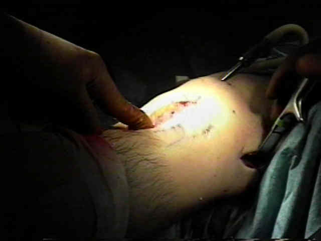
- notchplasty check:
- arthroscopic exam should show no impingement in full extension;
A biomechanical comparison of different surgical techniques of graft fixation for anterior cruciate ligament reconstruction.
The biomechanics of interference screw fixation of patellar tendon anterior cruciate ligament grafts.
Metal and biodegradable interference screws: comparison of failure strength.