

- Work Up for Scaphoid Fracture: (w/ discussion)
 - clinical differential diagnosis:
- distal radius frx
- transscaphoid perilunate dislocation:
- scaphoid impaction syndrome
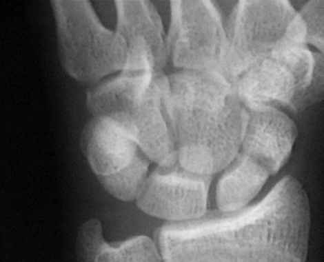
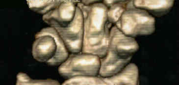
- radiographs and determination of stability (CT scan)
- non diagnostic radiograph (bone scan)
- tubercle frx
- transverse waist frx
- proximal pole frx
- treatment:
- non-displaced fractures
- casting of scaphoid frx
- percutaneous scaphoid fixation
- surgical treatment of displaced fracture (herbert screw fixation of scaphoid fractures):
- complications:
- nonunion of scaphoid (3.5 mm cannulated screw fixation)
- non union of proximal pole
- bone grafting technique
- avascular necrosis of the scaphoid
- SLAC or SNAC wrist
- degenerative disease of the STT joint:
- Degenerative changes at the scaphotrapezial joint following Herbert screw insertion: a radiographic study comparing patients with scaphoid fracture and primary hand arthritis.
- Discussion:
- surface of scaphoid is largely covered by articular cartilage, & only narrow area of its neck, & even smaller distal portion, are
accessible to blood vessels;
- frxs across scaphoid may destroy blood supply to its proximal part;
- scaphoid represents floor of anatomic snuff box;
- scaphoid spans both carpal rows and therefore has less mobility than other carpals;
- scaphoid is principal bony block to dorsiflexion of hand & wrist & is suscepible to frx during fall on outstretched hand;
- scaphoid (navicular): the most frequently fractured carpal bone (frx occurs in tubercle, waist, or proximal 1/3);
- biomechanics and scaphoid movement:
- scaphoid exerts flexion extension control over lunate and distal carpal row;
- ulnar side of the wrist exerts rotational control and stability;
- as wrist rotates from neutral to ulnar deviation, proxomal row dorsiflexes & x-ray profile of the scaphoid appears longer;
- in radial deviation, proximal carpal row volar flexes & scaphoid appears foreshortened;
- hence, ulnar deviation AP is necessary for visualization of scaphoid;
- because scaphoid crosses both proximal & distal carpal rows, excessive dorsiflexion causes it to be pinned between dorsal lip
of radius & palmar sling of the radial capitate ligament;
- scaphoid flexes with wrist flexion & extends with wrist extension, but it also flexes during radial deviation & extends
w/ ulnar deviation;
- these factors make immobilization of scaphoid fractures difficult;
- w/ scaphoid frx, distal scaphoid tends to flex, & proximal scaphoid extends with the proxmal carpal row;
- because of this, angulation occurs at frx site, which gaps open dorsally & gradually assumes a humpback deformity;
- mechanism of fracture:
- most injuries to wrist are sustained by a fall on outstretched hand;
- frx occurs w/ wrist is dorsiflexed & radially deviated;
- in this position, proximal pole of schaphoid is held by radius & radioscaphocapitate ligament, while distal pole of bone is
carried dorsally by trapeziocapitate complex;
- radioscaphoid ligament is relaxed by & radial deviation & cannot alleviate tensile stresses accumulating on radiovolar
aspect of scaphoid:
- radioscaphoid ligament:
- inserts onto tuberosity of scphoid & is radial expansion of radiocapitate ligament which courses over palmar concavity
of scaphoid proximal to tuberosity before inserting on palmar aspect of capitate;
- forms a fulcrum over which scaphoid rotates;
- incidence:
- 1 out of 100,000 people per year;
- ref: Incidence Estimates and Demographics of Scaphoid Fracture in the U.S. Population
- Pediatric Scaphoid Fracture:
- forms from enchondral ossification
- forms in males between ages 5-15
- forms in forms in females 4-13 years;
- non operative treatment is usually indicated;
- references:
- Pediatric fractures of the carpal scaphoid: a retrospective clinical and radiological study
- Pediatric scaphoid nonunion