

- See:
- Scaphoid Reconstruction for Nonunion
- Vascular Anatomy of Scaphoid:
- Discussion:
- may be associated w/ longstanding nonunion of proximal pole fractures, especially when associated w/ previous surgery;
- AVN of scaphoid is often difficult to diagnose radiographically and therefore it is usually necessary to assess vascularity of the
proximal pole at the time of surgery;
- absence of punctate bleeding in the proximal fragment (after debridement) is the best indicator of AVN;
- following debridement, punctate bleeding should be present from the surface of the scaphoid while the tourniquet remains elevated;
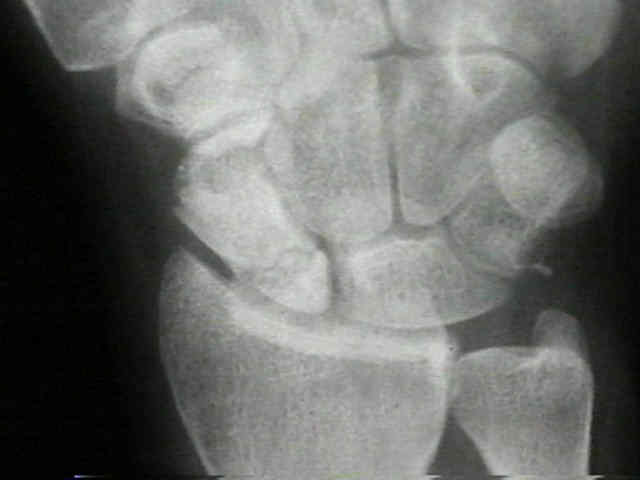
- Radiographic Findings:
- plain radiographs tend to underestimate the presence of AVN (as compared to intraoperative bleeding or MRI);
- proximal fragment may have:
- ground-glass appearance or increased bone density;
- loss of trabecular pattern;
- cystic changes;
- subchondral collapse and fragmentation;
- radiographic classification:
- stage 0: none;
- stage 1: patchy areas of radiodensity of proximal pole;
- stage 2: involvement of entire proximal pole;
- stage 3: AVN w/ carpal collapse;
- MRI:
- evolving role;
- Treatment:
- Four Corner Fusion:
Avascular necrosis after scaphoid fracture: a correlation of magnetic resonance imaging and histology.