- Discussion: 
- a chronic progressive disease osteoblasts and osteoclasts which results in abnormal bone remodeling;
- etiology, staging, and pathogenesis
- prevalence:
- uncommon in pts under age of 55 yrs but relatively common in later life, occurring in 3-4% of persons over 55 yrs &
occurs in 10% in pts over age 80;
- reference:
- A comparison of the amount of vascularity in pagetic and normal human bone.
- Clinical Presentation:
- frequently is asymptomatic and diagnosed incidentally on x-rays obtained for other purposes;
- may be localized to only one or several bones;
- relative frequency of involvement: pelvis, femur, skull, tibia, and least often involved is the vertebrae;
- wt bearing bones:
- may present w/ pain, increasing deformity, Pathologic Frx,
- local pain & tenderness over the affected bone are frequent.
- one or many bones may be involved.
- there may be night pain unrelated to change of position
- ref: Images in Clinical Medicine. Saber Tibia in Paget's Disease of Bone
- spine:
- neural compression may be the presenting problems;
- skull:
- headache and deafness due to skull involvement;
- hat size may increase;
- skull radiographs may reveal osteoporosis circumscripta which is a lytic lesion seen in the early stages of Paget's disease;
- ref: Images in Clinical Medicine. “Cotton Wool” Appearance of Paget's Disease
- cardiovascular:
- may have high output failure and high pulse due to increase blood flow thru Pagetoid bone;
- Histology:
- demonstrates excess osteoclasts resorptive activity; (occurs predominantly in the early resorptive phase);
- bone marrow is replaced by fibrous tissue and disorganized trabeculae;
- paratrabecular fibrosis;
- irregular - highly celluar lamellar bone w/ irregular cement lines;
- woven bone with osteoblastic rimming;
- occasional prominent cement lines.
- Radiographs:
- aggressive bone resorption;
- lytic lesions w/ sharp borders that destroy the cortex;
- new bone formation and sclerosis which causes thickening of the cortex and course trabeculae;
- cortices will appear thickened and trabeculae will appear coarse;
- skull radiographs may reveal osteoporosis circumscripta which is a lytic lesion seen in the early stages of Paget's disease;
- ref: Images in clinical medicine. Saber tibia in Paget's disease of bone.
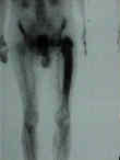
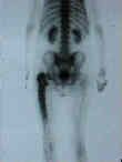
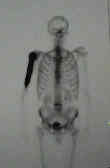
- Bone Scan: 


- Lab Dx:
- there is a marked increase in alk phos, which reflects increase in bone formation (w/ symptoms alk phos is 2x normal);
- serum Ca & Phos, & acid phosphatase are usually normal (unless patient has been immobilized in which case
hypercalemia may occur);
- uric acid level may be increased;
- urinary hydroxyproline:
- increased serum levels of hydroxyproline levels & increased urinary levels of hydroxyproline;
- elevated urinary hydroxyproline is a reflection of bone lysis and may also occur in RA, hyperparathyroidism, and OM;
- urinary pyridinium / pyridinoline peptides can also be used as markers of increased activity;
- Treatment:
- indications for treatment:
- use of antipagetic medications prior to elective surgery is recommended;
- goal is to reduce hypervascularity assoc w/ active disease (3x alk phos) which will reduce the amount of blood loss at operation;
- to reduce bone pain (probably the most common complaint), excessive warmth over bones, headache, low back pain, &
neural compression;
- pain due to secondary arthritis from pagetic bone may or may not respond to antipagetic treatment;
- presence of moderately active asymptomatic dz than normal at sites where potential problems or complications exists (wt
bearing joints, spine, etc.);
- presence of monstotic dz of a tibia or femur with minimal elevation of alk phos are also treatment candidates;
- medications:
- bisphosonates:
- first line agent (low doses interfere w/ bone resorption where as high doses impair bone mineralization);
- fosamax (alendronate): 40 mg/day (first line agent);
- pamidronate: second line agent given IV (if fosamax is not tolerated);
- etidronate
- it is an oral agent and is less expensive than either of the calcitonins;
- while side effects are < calcitonin, the efficacy is also less;
- plicamycin
- calcitonin
- w/ extensive lytic disease in a wt bearing bone, calcitonin is the drug of choice esp with prior to elective surgery;
- indications: neurologic compression, given to patients prior to orthopaedic surgery or for pathologic frx, pain
or deformity;
- symptomatic benefit may be apparent in a few weeks;
- alkaline phosphatase will decrease after 3-6 months;
- major drawback is vomiting in 10-20% of patients;
- TKR in Paget's disease:
- see total knee replacement menu;
- tibial and femoral bowing associated with Paget disease often necessitates extramedullary cutting guides;
- arthritic knee pain must be differentiated from pain caused by Paget disease;
- arthritic knee is confirmed as source of pain if that pain is relieved after intra-articular local anesthetic injection or ineffectively
relieved following diphosphonate or calcitonin therapy;
- ref: Total knee arthroplasty for osteoarthrosis in patients who have Paget disease of bone at the knee.
- Paget's Osteosarcoma:
- references:
- Survival and management considerations in postirradiation osteosarcoma and Paget's osteosarcoma.
- Paget's Osteosarcoma - no Cure in Sight
References:
- On a form of chronic inflammation of bones. (Osteitis deformans).
- Osteolytic Paget's disease. Recognition and risks of biopsy.
- Long-term follow-up observations on treatment in Paget's disease of bone.
- Efficacious management with aminobisphosphonate (APD) in Paget's disease of bone.
- Experimental basis for the use of bisphosphonates in Paget's disease of bone.
- New modes of administration of salmon calcitonin in Paget's disease. Nasal spray and suppository.
- Combination drug therapy in treatment of Paget's disease of bone: clinical and metabolic response.
- Intranasal and intramuscular human calcitonin in female osteoporosis and in Paget's disease of bones: a pilot study.
- Vitamin D status in Paget's bone disease. Effects of calcitonin therapy.
- Metabolic consequences of bone turnover in Paget's disease of bone.
- Skeletal distribution and biochemical parameters of Paget's disease.
- Dynamic radiologic patterns of Paget's disease of bone.
- Total hip replacement for Paget's disease of the hip.
- Osteotomy for tibia vara in Paget's disease under cover of calcitonin.
- Osteolytic form of Paget's disease. Differential diagnosis and pathogenesis.
- Paget disease of the calcaneus: findings on MR imaging..
- Corrective Osteotomy for Deformity in Paget Disease.
Other links:
- Paget's Disease of the Bone from the Orthopaedic Care Textbook
- Paget's Sarcoma from the Orthopaedic Care Textbook

