

- See:
- Femoral and Tibial Traction Pins
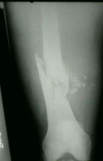
- Supracondylar Femoral Fractures
- Discussion:
- failure of the nail to fill the canal of either the proximal or distal fragment, may lead to postoperative instability;
- distal fractures within 10 cm of the joint line often can be treated successfully with standard IM nailing;
- Traction Pins:
- when treating distal fractures, the knee must be flexed and a distal femoral traction pin must be inserted;
- flexed knee position releaxes the posterior knee capsule and the gastrocnemius muscle, thereby avoiding
a hyperextension deformity at the fracture, which can prevent fracture reduction;
- femoral traction pin is inserted anteriorly just proximal to the adductor tubercle, and is placed from the inframedial to
superolateral side to pull the fracture out of valgus angulation;
- because the distal femoral traction pin needs to be placed very distal and anterior, consider the use of fluoroscopy to
avoid penetration into the knee joint;
- note: in the lateral position, the weight of the leg produces a valgus angulation at the fracture site;
- if the deformity is not corrected during the insertion of the nail, the nail will be driven into the medial femoral condyle, and
a valgus deformity will result;
- finite element analysis of interlocking nails, have revealed that if a frx is located w/in 5 cm of this hole, stresses are generated
in the nail above its endurance limit;
- Reduction:
- frx of distal third of the shaft pose a special reduction problem;
- in supine position, the distal fragment angulates posteroirly and must be supported with a crutch;
- in lateral position, the dstal fragment sags into valgus angulation;
- Reaming:
- faster union may be achieved in distal femoral shaft fractures which have been reamed vs. those that have not been reamed;
- in these frxs, reaming allows insertion of a larger nail, which allows more rigid fixation between the implant and the bone;
- IM Nail Technical Considerations:
- IM nails for supracondylar fractures:
- tend to sag into a valgus position;
- distal purchase of the nail is critical for stability;
- major loading of this region of femur, along w/ inadequacy of endosteal purchase on distal frag, results also in a higher non union
rate w/ interlocking nails than is seen in midshaft fractures;
- thus, the cancellous bone is not reamed;
- distal third frx especially require minimal 1.0 to 1.5 mm overreaming of proximal fragment to accomodate the variable degree of
anterior femoral bow that might be present;
- nail is driven thru old epiphyseal scar to level of intercondylar notch, hence an appropriately sized nail is extremely important;
- after nail has been driven a few mm across frx, traction may be decr sufficiently to allow impaction of frx as nail is driven distally;
- frx angulation is also possible intraoperatively if the nail is driven eccentrically out of alignment w/ longitudinal axis of the canal;
- w/ distal fractures the nail may be driven into the medial or lateral condyles resulting in either a valgus or varus deformity;
- if full correction of this problem is not achieved before guide pin insertion, the nail may be driven into the medial femoral
condyle, resulting in a valgus deformity;
- guide pin should be aimed directly at intercondylar notch on AP view of femur before reaming and nailing of distal fragment;
- reaming of distal fragment down to anticipated distal tip of nail is unnecessary and may comprimise the purchase of nail on
cancellous bone of the distal third of the shaft;
- Length of Nail:
- distal end of medullary nail should be at superior pole of patella in isthmal level fractures &, for more distal fractures, just
proximal (approx 3 cm) to the intercondylar notch;
- make allowance for the slight overdistraction at the fracture site;
- to prevent problems w/ protrussion into gluteal muscles, it should not extend above the greater trochanter;
- in some cases further impaction occassionally occurs when severly comminuted fractures are later dynamized;
- this should be considered to prevent later nail migration into the knee or nail protrussion out of the proximal femur
- References:
Nails or plates for fracture of the distal femur?