

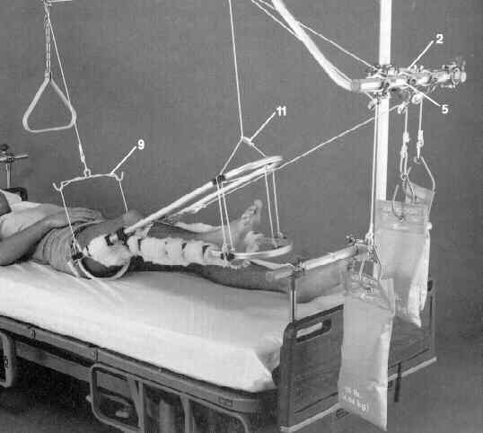
- skeletal traction can be applied under sedation & local anesthesia;
- Distal Femoral Pins:
- inserted on medial side to avoid injury to femoral artery on pin exist;
- flex the knee and thigh on several folded sheets inorder to facilitate pin insertion from the opposite side of the table (medial to
lateral) and to facilitate obtaining a lateral radiographic view;
- entry site is just proximal adductor tubercle (proximal to medial epicondyle and/or growth plate);
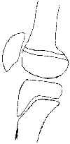
- distal pin placement risks entering joint at intercondylar notch;
- more proximal pin insertion risks injury to femoral artery at Hunter's canal;
- flex knee to 90 deg:
- traction pin must be applied w/ knee at 90 deg of flexion;
- if leg is in extension while pin is inserted, it will later be difficult to flex the knee because the pin is bound by the IT band;
- as the short longitudinal incision is made, turn the knife 90 deg (once it is buried under the skin) in order to make a small
transverse nick in the IT band;
- place pin perpendiulcar to knee joint, rather than perpendicular to femoral shaft;
- Proximal Tibial Pins: 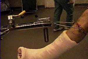
- contraindications:
- ligament injury to ipsilateral knee;
- should never be used in children;
- may cause recurvatum injury due to damage of tibial physis;
- pins are inserted from lateral side to avoid damaging peroneal nerve;
- pin insertion:
- proper insertion site: 2.5 cm posterior to & 2.5 cm distal to tibial tubercle;
- landmark is to place pin one to two fingerbreaths below tibial tuberosity in the midportion of the tibia;
- proximal pin placement, places it thru too much cancellous bone, which is weaker;
- distal pin placement, while in stronger cortical bone, risks damage to peroneal nerve as it passes anterior after it passes
around fibular neck;
- make a transverse skin incision about 1 cm in length, placed about 3 cm below lesser tuberosity;
- the most common mistake is to make the incision too anterior, which causes the skin to bunch up posteriorly