

- Discussion:
- advantages of femoral shaft reaming:
- avoids potential mis-sizing of nail diameter (which can occur when non-reamed nails are inserted);
- reaming may carry bone graft particulate into the fracture site;
- faster union may be achieved in distal femoral shaft fractures which have been reamed vs. those that have not been reamed;
- in these frxs, reaming allows insertion of a larger nail, which allows more rigid fixation between the implant and the bone;
- in the study by Clatworthy MG, et al. (1998), reamed vs unreamed femoral nails were compared in a randomized prospective trial;
- frxs in the unreamed group were slower to unite, required more secondary procedures, and had more implant failures;
- the authors recommended insertion of at least 12 mm nails in females and 13 mm nails in males;
- disadvantages of reaming:
- potential for fat embolism syndrome
- referenceL Reamed versus unreamed femoral nails. A randomised prospective trial.
- Guide Wire Insertion:
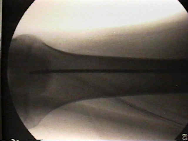
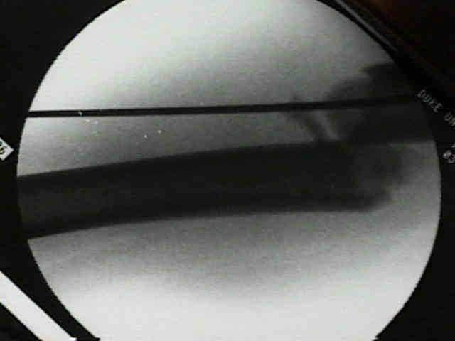
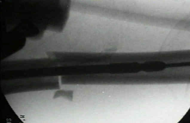
- at frx site, wire is advanced few mm at time w/ flouro to confirm IM passage;
- unrecognized advancement of the guide wire thru soft tissues may produce unrecognized neurologic or vascular damage;
- distally, guide wire, is advanced centrally down distal fragment & is seated against subchondral bone;
- w/ distal frxs, the guide wire can easily be inadvertantly drawn back across the fracture site during reaming;
- to prevent this, clamp & stabilize guide wire during reaming;
- hazards:
- once the guide wire has been passed, it is important to check its position on two views;
- Optimal Reaming Characteristics:
- reamer should have a narrow shaft w/ deep blades and hollow contruct;
- in the study by Mousavi M, et al, the authors assessed the influence of driving speed and revolution rate per minute of
two reamers on femoral IM pressure increases and fat intravasation;
- AO and Howmedica reamers were tested in four groups with different combinations of driving speed and revolution rate per minute
in both femurs in a sheep model.
- 24 animals were exposed to hemorrhagic shock after midshaft osteotomy and were resuscitated before reaming of both femoral shafts;
- controlled reaming was performed at 15 and 50 mm/second driving speed with 150 and 450 revolutions per minute.
- fat intravasation and IM pressure were measured by transesophageal echocardiography, Gurd test, and a piezoelectric gauge, respectively.
- low driving speed and high revolutions per minute with the smaller cored reamer led to lower intramedullary pressure changes.
- same reaming parameters led to greater pulmonary stress during surgery of the second side;
- primary reamed intramedullary nailing should be done after resuscitation at a low driving speed and high revolutions per minute
with a smaller cored reamer to minimize the risk of pulmonary dysfunction;
- ref: Influence of Controlled Reaming on Fat Intravasation After Femoral Osteotomy in Sheep.
- Reaming:
- inorder to avoid FES, ream slowly w/ gentle pressure;
- reaming should progress in 1 mm increments until the cortex is reached and then in 0.5 mm increments thereafter;
- usually ream one mm larger once additional chatter is encountered;
- w/ stiffer closed section interlocking nails, overreaming by 1.5 mm is recommended to allows easier nail insertion;
- design changes have affected the biomechanics of IM nails;
- softer nails made of titanium w/ anatomic fit require less over reaming (only 1 mm);
- relatively stiff nails (stainless steel) w/ large radius of curvature may require overreaming of upto 2 mm;
- run each reamer back and forth several times along the meduallar canal until it passes easily;
- prevent eccentric reaming of the cortex, by making sure that guide pin, in the AP and lateral image views is centered in distal metaphysis;
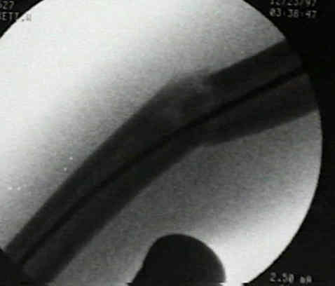
- note that even after the guide wire has reduced the femoral shaft fracture, there will often be a "step off" on the lateral view, which
can lead to eccentric reaming;
- this is often due to hamstring tightness, which in the case of retrograde nailing, can easily be relieved by flexing the knee with a bump;
- ref: “Push-past” Reaming as a Reduction Aid With Intramedullary Nailing of Metadiaphyseal and Diaphyseal Femoral Shaft Fractures
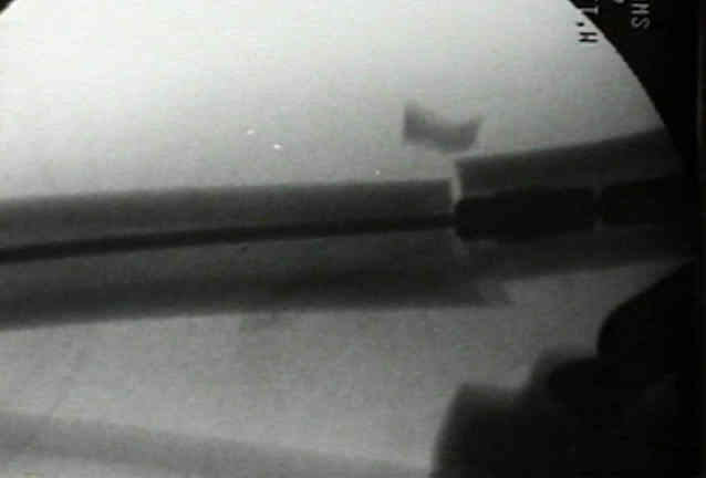
- 3 mm guide pin is exchanged for 4 mm guide pin, & nail is inserted;
- once nail has entered distal fragment, rotatory alignment of limb is checked & traction is adjusted to restore nl femoral length;
- distal 1/3 fractures:
- these frx especially require at least 1.0 to 1.5 mm of over reaming of proximal fragment to accommodate variable degree of anterior femoral bowing;
- Nail Width:
- generally the width of the nail will be 2 mm greater than the isthmal medullary width measured on the AP film
- Hazards:
- incarcerated reamer:
- can be extricated by withdrawing ball tip of guide wire against reamer & gently disimpacting the guide wire w/ a vise grip & mallet;
- conventional spring type flexible reamers cannot be reversed or they will unwind within medullary canal;
- as noted by Duwelius, et al (1997), IM reaming tends to cause a transient increase in pulmonary vascular resistance;
- fat embolism syndrome:
- may occur as a result of increased intramedullary pressure and resultant fatty emboli;
- as noted by Bosse, et al (1997), IM nailing w/ reaming in multiply injured patients did not increase in incidence of FES or ARDS
as compared to femoral shaft fractures treated w/ plate fixation;
- always attempt to use a cannulated nail (if possible) in order to reduce marrow extravasation during nail insertion;
- references:
- The effects of femoral intramedullary reaming on pulmonary function in a sheep lung model.
- Adult respiratory distress syndrome, pneumonia, and mortality following thoracic injury and a femoral fracture treated either with intramedullary nailing with reaming or with a plate. A comparative study.
- Early unreamed intramedullary nailing of femoral fractures is safe in patients with severe thoracic trauma.
- Fat extravasation due to unreamed and experimentally reamed intramedullary nailing of the sheep femur.
- Fatal pulmonary embolization after reaming of the femoral medullary cavity in sclerosing osteomyelitis: a case report.
- Pulmonary damage after intramedullary femoral nailing in traumatized sheep--is there an effect from different nailing methods?
- Femoral canal reaming in the polytrauma patient with chest injury. A clinical perspective.
- Reamed versus unreamed intramedullary nailing of the femur: comparison of the rate of ARDS in multiple injured patients.
- Femur Fractures and Lung Complications: A Prospective Randomized Study of Reaming.
Curvature of the femur and the proximal entry point for an intramedullary rod.
Intramedullary nailing and reaming for delayed union or nonunion of the femoral shaft. A report of 105 consecutive cases.
Reamed versus unreamed femoral nails. A randomised prospective trial.
Nonunion Following Intramedullary Nailing of the Femur with and without Reaming. Results of a Multicenter Randomized Clinical Trial.
Intramedullary pressure increase and increase in cortical temp during reaming of femoral cavity: the effect of draining the medullary contents before reaming.
The thermal effects of intramedullary reaming.
..............................................................................................................................................................................................................................................................................................................................................................