

- See:
- septic arthritis
- general orders and treatment
- Discussion:
- septic arthritis of the hip is a surgical emergency;
- it is essential that dx be made ASAP to prevent joint damage;
- if any pus is obtained from arthrotomy, then immediate arthrotomy is indicated, regardless of the Graim Stain results;
- w/ younger child, more pressing is need because of higher risk of permanent disability;
- pathophysiology:
- in infrants, septic arthritis may occur from propagation of adjacecent proximal femoral osteomyelitis; (see osteomyelitis in the infants);
- differential dx:
- note that because septic arthritis of the hip requires prompt surgical drainage, it is essential to make a prompt correct diagnosis;
- Kocher criteria: (for child with painful hip)
- includes: non-weight-bearing on affect side, sed rate greater than 40 mm/hr, fever, and a WBC count of >12,000 mm3;
- when 4/4 criteria are met, there is a 99% chance that the child has septic arthritis;
- when 3/4 criteria are met, there is a 93% chance of septic arthritis;
- when 2/4 criteria are met, there is a 40% chance of septic arthritis;
- when 1/4 criteria are met, there is a 3% chance of septic arthritis;
- references:
- Differentiating Between Septic Arthritis and Transient Synovitis of the Hip in Children: An Evidence-Based Clinical Prediction Algorithm.
- A guideline for treatment of septic arthritis in children: efficacy in improving care and effect on outcome of septic arthritis of the hip.
- Differentiation between septic arthritis and transient synovitis of the hip in children with clinical prediction algorithms.
- Associated Conditions:
- hematogenous osteomyelitis:
- septic arthritis is both more common & more often assoc w/ metaphyseal osteomyelitis in neonates than in older children;
- approximately 20% of infants with septic arthritis have adjacent osteomyelitis, but more than half of neonates w/ septic
arthritis may have concomitant osteomyelitis;
- epiphyseal plate prevents infection from entering joint space in older children but apparently does not act as a barrier in infants;
- these transphyseal blood vessels are thought to disappear by age 6 mo, but the exact age of this transition remains unclear;
- joint infection secondary to osteomyelitis may occur in shoulder and hip as result of the synovial membrane inserting distally to epiphysis, allowing
bacteria to spread directly from the metaphysis to joint space;
- only metaphysis of shoulder, hip, radial head, and ankle remain intracapsular during early childhood;
- the hip joint seems especially prone to sepsis from adjacent osteomyelitis;
- these synovial reflections over the metaphyseal bone decrease with age;
- note that in cases of osteomyelitis of the femoral neck some consideration of femoral neck drilling should be made
noting that there is some evidence that this may prevent progression to septic arthritis;
- references:
- The arterial supply of the developing proximal end of the human femur.
- Acute hematogenous osteomyelitis of the neck of the femur in children treated with drilling.
- Exam:
- child w/ septic arthritis of hip usually complains of pain in groin area that occasionally radiates down the medial side of thigh;
- pain tends to increase gradually, developing to an excruciating level, and may be accompanied by spasm of the hip muscles;
- hip is usually kept in a position of flexion and external rotation & affected hip will have decreased internal rotation compared to the normal hip;
- patient resists all attempts to move hip;
- palpate the SI joint for local tenderness;
- Labs:
- synovial fluid exam (total cell count)
- C-reactive protein:
- sed rate
- at initial presentation the sed rate is usually greater than 20 mm but may increase above 90 mm after 2-3 days;
- allows monitoring of the response to treatment of pt w/ septic hip;
- serial determinations of the ESR is helpful in monitoring the course of hematogenous septic arthritis, as a persistently elevated ESR has been
found to be a reliable indicator of continued activity of the disease;
- aspiration:
- see technique of aspiration
- it is helpful to use GEA in children as well as flouroscopy;
- if aspiration is performed in the OR, open drainage can be carried out if the aspirate yields pus or cloudy fluid;
- aspiration of joint should be done in every case of suspected septic arthritis & esp in cases of suspected septic arthritis of hip;
- bacteriology:
- neonates:
- ages from 6 months to 2 years:
- age greater than 2 years:
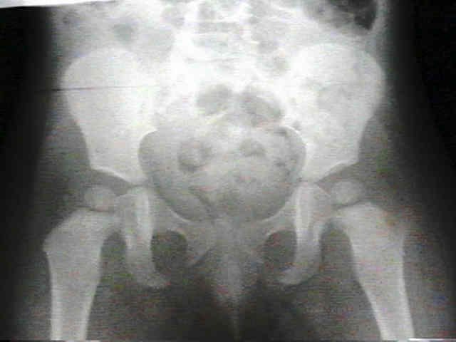
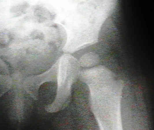
- Radiographs:
- x-rays early in course of disease may show evidence of soft-tissue swelling or hip joint capsular distension (w/ widening of the
joint space or even subluxation);
- look for radiographic changes in the proximal femoral metaphysis that might indicated adjacent osteomyelitis;
- bone scan:
- many feel that bone scans are a poor choice to rule out adjacent osteomyelitis in infants < 6 months (w/ negative studies in up to 60% of cases of OM):
- gallium is more sensitive but exposes the child to a higher dose of radiation;
- indium labeled scans are not often used in the diagnosis of septic hip because of the relatively large amount of blood specimen required
and the delays in obtaining the results of this study;
- three phase:
- in the hip and shoulder, increased intracapsular pressure may cause ischemia of the femoral or humeral head, which manifests
as increased signal on the delayed bone images;
- MRI
- may be the most useful test to distinguish proximal femoral osteomyelitis from septic arthritis of the hip;
- in one anecdotal case, an aspiration was attempted in a suspected case of septic arthritis but no fluid was obtained;
- an MRI was obtained which revealed a hip effusion and a repeat aspiration yielded pus;
- in some situations, MRI will diagnose other conditions such as psoas abscess or other extra-articular cases of sepsis;
- ref: The Role of Pelvic Magnetic Resonance in Evaluating Nonhip Sources of Infection in Children With Acute Nontraumatic Hip Pain.
- Surgical Drainage:
- septic hips are drained via an anterior approach because this avoids femoral blood supply and allows easy access to
proximal femoral metaphysis for inspection and possible drilling;
- make oblique anterior incision beginning 1 cm below ASIS which is then extended inferiorly and medially;
- separate the TFL and vastus lateralis on the lateral side and the sartorius on the medial side;
- identify the lateral border of the rectus, and reflect it medially;
- 1 square cm anterior capsulectomy is made;
- joint thoroughly lavaged with saline;
- avoid drilling a hole in the neck of femur: either decompression into the joint has already occurred, or primary osteomyelitis of metaphysis of
proximal femur may be treated by antibiotic therapy, which should be commenced immediately after fluid and synovium are
obtained for culture;
- if the joint is grossly unstable after decompression, an abduction plaster or spica cast is applied (otherwise, no immobilization is used);
- leave capsule open, and close skin over drains (drain should be removed in 24 to 48 hours);
- cefuroxime is the initial systemic antibiotic of choice; it may be changed according to results of cultures and sensitivity studies;
- Complications and Management:
- septic arthritis of hip may lead to dislocation, subluxation, dysplasia, coxa vara, coxa breva, absence of head & neck of femur, and postinfectious arthritis;
- numerous reconstructive procedures, such as open reduction, pelvic osteotomy, femoral osteotomy, and greater trochanteric
arthroplasty, have been advocated to treat the residual deformity;
- however, whether these procedures performed at any stage are less favorable than natural history of the deformity;
- hip dislocation:
- infantile hip sepsis causes destruction of the femoral head patient and led to the high-riding dislocation and failure of acetabular development.
- leg length descrepancy:
- the proximal femoral epiphysis may be destroyed by the septic process, with a resultant limb length descrepancy of 3-4 inches;
- femoral lengthening should not be attempted if hip stability is not present;
- if an acetabulum is present, surgical reduction w/ trochanteric arthroplasty and pelvic osteotomies may be successful, but surgical reconstruction
is less successful than closed treatment of the hip, use of shoe lift, and later distal femoral epiphysiodesis to treat leg length difference
Suppurative arthritis of the hip in children.
Long-term follow-up of infantile hip sepsis.
Sequelae and reconstruction after septic arthritis of the hip in infants.
Acute septic arthritis of the hip joint in infancy and childhood.
Pyogenic arthritis associated with adjacent osteomyelitis: identification of the sequela-prone child.
Significance of laboratory and radiologic findings for differentiating between septic arthritis and transient synovitis of the hip.
A clinical practice guideline for treatment of septic arthritis in children: efficacy in improving process of care and effect on outcome of septic arthritis of the hip.
Treatment of Septic Arthritis of the Hip Joint by Repeated Ultrasound-guided Aspirations.