- see distal radius fracture menu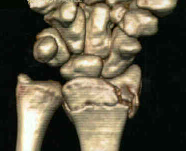 (volar Barton's frx)
- orif of distal radius fractures
- Surgical Approach:
- standard volar approach:
- most indicated for simple extra articular fractures and when there is absence of ulnar comminution / displacement:
- incision starts at thenar crease of palm, curves toward middle of forearm, w/ transverse segment as it crosses volar flexion crease of wrist;
- dissection goes through FCR tendon tendon sheath and then the FCR is then retracted ulnarly and the incision is continued thru dorsal sheath, down to FCR and radial artery - radial artery need not be exposed;
- it is simply retracted radially and protected by the surrounding soft tissues;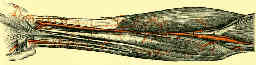
- controversies: carpal tunnel syndrome
- w/ severe CTS symptoms use standard open approach to palmar cutaneous branch is avoided);
- ref: Routine carpal tunnel release not needed when plating distal radius fractures
- pronator quadratus muscle is taken down from its radial origin to expose underlying fracture;
- this will need to be repaired at the end of the case;
- since repair of the pronator can be difficult, it can be tied down to the radial edge of the butress plate;
- avoid excessive retraction of the median nerve;
- ulnar sided comminution / displacement: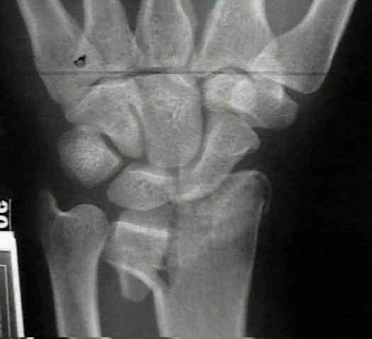
- if predominate comminution or displacement is on ulnar side of volar radius, then consider an extended carpal tunnel incision which crosses proximal wrist crease obliquely and which then courses further proximally ulnar to PL tendon;
- a wide exposure is needed inorder to achieve adequate exposure of the volar radius;
- approach radius on ulnar side of flexor tendons inorder to avoid trauma to median nerve and its palmar cutaneous branch;
- excessive ulnar retraction of the median nerve (w/ a standard Henry approach) may lead to RSD;
- Reduction:
- optimally, it is best to achieve reduction prior to plate application;
- reduction is achieved with supination and dorsiflexion over rolled towel, and is confirmed w/ flouroscopy;
- reduction is held w/ K wires inserted from volar to dorsal, which are oriented to allow holes of the T plate to slide down wire;
- volar capsule should not be opened for inspection as this violates radioscaphocapitate and radioscapholunate ligaments;
- articular reconstruction is supported by autogenous cancellous-bone graft, as well as a small buttress plate;
- in some cases, with initial fixation of the distal plate screws, the proximal end of the plate will be proud over the cortex;
- with opposition of the this end of the plate over the cortex, there will be increase volar tilt, which can improve the reduction;
- Internal Fixation:
- (Synthes Distal Radius Plates)
- distal end of plate should be placed far enough proximally to avoid insertion of screws into articular surface;
- consider insertion of ulnar screws first to ensure that there is no joint tresspass (radial screws obstruct view of ulnar screws);
- bend plate to comform to the normal configuration of the radius (contour plate around the radial styloid), esp in smaller patients;
- screws placed in diaphyseal bone will act as a butress for distal fragment;
- avoidance of hardware complications:
- radiographs:
- 11 degrees tilt PA : perfect articular PA view should correctly demonstrate whether screws have penetrated the joint;
- 22 degrees tilt lateral views
- slightly less tilt (15-23 deg) will correctly visualize the ulnar facet, and 30 deg tilt will correctly visualize the radial styloid;
- references:
- Fluoroscopic Evaluation of Intra-Articular Screw Placement During Locked Volar Plating of the Distal Radius: A Cadaveric Study
- Use of Articular Wrist Views to Assess Intra-Articular Screw Penetration in Surgical Fixation of Distal Radius Fractures
- Rotational fluoroscopy assists in detection of intra-articular screw penetration during volar plating of the distal radius
- Anatomic tilt x-rays of the distal radius: an ex vivo analysis of surgical fixation.
- Lateral tilt wrist radiograph using the contralateral hand to position the wrist after volar plating of distal radius fractures
- Tangential views of the articular surface of the distal radius-aid to open reduction and internal fixation of fractures
- Radiographic evaluation of dorsal screw penetration after volar fixed-angle plating of the distal radius: a cadaveric study.
- Fluoroscopic evaluation of intra-articular screw placement during locked volar plating of the distal radius: a cadaveric study.
- pronator quadratus repair:
- The Effects of Pronator Quadratus Repair on Outcomes After Volar Plating of Distal Radius Fractures.
- Assessment of pronator quadratus repair integrity following volar plate fixation for distal radius fractures: a prospective clinical cohort study.
- Complications of ORIF
- EPL tendon injuries:
- frequent reports of extensor tendonitis or rupture from dorsally and volarly applied hardware;
- causes:
- postreduction bone spurs
- dorsal gapping at the fracture site
- prominent screw tips
- Flexor Tendon Injuries:
- watershed line:
- transverse ridge situated within 2 mm of radial joint line on ulnar side of radius, and 10-15 mm from the articular surface on radial side of bone;
- flexor tendons are separated from the cortex by the pronator quadratus;
- plates applied distal to watershed line may come in direct contact with the flexor tendons;
- references:
- Flexor Tendon Injuries Following Locked Volar Plating of Distal Radius Fractures
- Perforation of the third extensor compartment by the drill bit during palmar plating of the distal radius.
- Radiographic evaluation of dorsal screw penetration after volar fixed-angle plating of the distal radius: a cadaveric study.
- Loss of Reduction:
- Rozental and Blazar reported on 2 of 41 patients who developed dorsal collapse of the fracture resulting in excessive dorsal tilt
- 50 distal radius fractures treated with volar plate fixation (49 patients), 4 patients experienced angular instability
- references:
- Functional outcome and complications after volar plating for dorsally displaced, unstable fractures of the distal radius.
- Osteosynthesis of distal radial fractures with a volar locking screw plate system.
- Operative management of distal radial fractures with 2.4-millimeter locking plates: a multicenter prospective case series. Surgical technique.
- References
Surgical treatment of fractures of the distal radius with plates: a comparison of palmar and dorsal plate position
Combined Dorsal and Volar Plate Fixation of Complex Fractures of the Distal Part of the Radius.
Loss of Fixation of the Volar Lunate Facet Fragment in Fractures of the Distal Part of the Radius.
Complications following internal fixation of unstable distal radius fracture with a palmar locking-plate.
A comparative study of clinical and radiologic outcomes of unstable colles type distal radius fractures in patients older than 70 years: nonoperative treatment versus volar locking plating.
Functional outcome of unstable distal radius fractures: ORIF with a volar fixed-angle tine plate versus external fixation.
Volar fixation of dorsally displaced distal radius fractures using the 2.4-mm locking compression plates.
Functional outcome and complications after volar plating for dorsally displaced, unstable fractures of the distal radius.
Operative management of distal radial fractures with 2.4-millimeter locking plates. A multicenter prospective case series.
Prospective study of distal radius fractures treated with a volar locking plate system
A randomized prospective study on the treatment of intra-articular distal radius fractures: open reduction and internal fixation with dorsal plating versus mini open reduction, percutaneous fixation, and external fixation.
Year Book: Fractures of the Lower End of the Radius Anteriorly Displaced Treated by Plating.
Internal fixation of volar-displaced distal radial fractures.
Anterior Margin Articular Fractures of the Distal Radius.
Barton's and Smith's fractures.
Volar fixed-angle plating of distal radius extension fractures: influence of plate position on secondary loss of reduction--a biomechanic study in a cadaveric model.
Treatment of Unstable Distal Radial Fractures with the Volar Locking Plating System. Surgical Technique
Volar approach to distal radius fractures.
Minimally invasive plate osteosynthesis of distal radius fractures using a pronator sparing approach.
Screw placement may eliminate iatrogenic extensor tendon-related complications
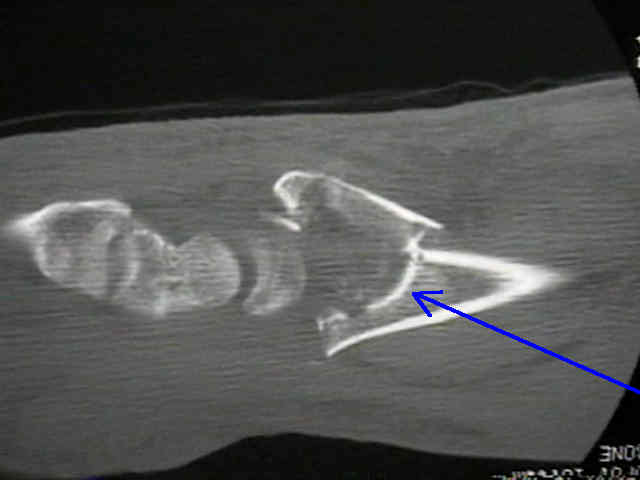
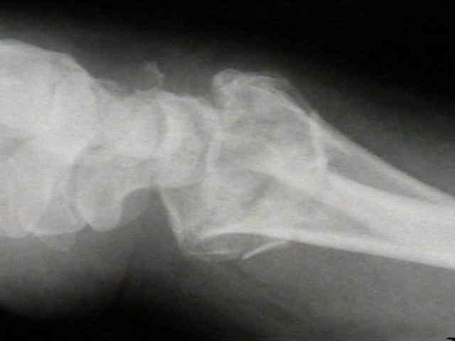
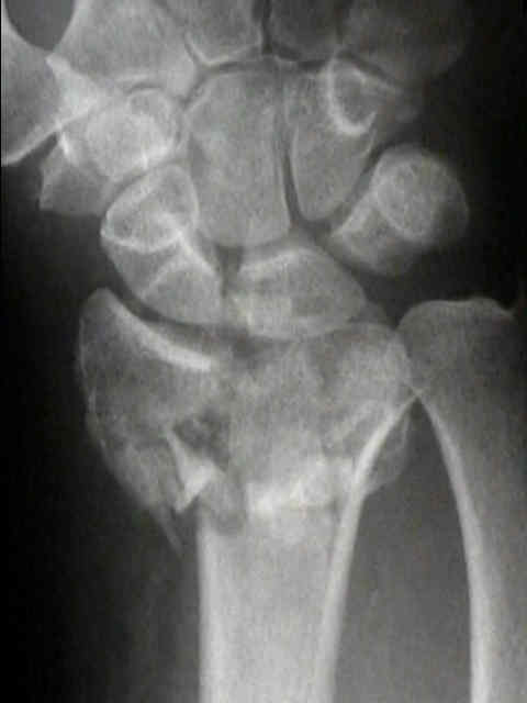
- Case Example: