

- See:
- Radiology of the Wrist
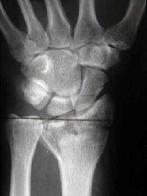
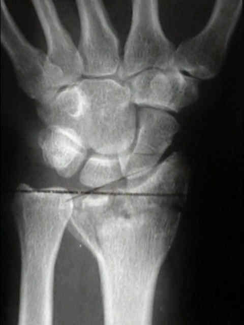
- Distal Radius Fx
- Discussion:
- radiographic measurements:
- articular step off: most important determinant of outcome;
- radial shortening second most determinant of outcome;
- dorsal angulation
- radial inclination
- unstable vs stable frx;
- stable frx are usually extra articular w/ mild to moderate displacement, & when reduced do not redisplace to original deformity;
- in stable extra-articular fractures, there will often be frx extension into the DRUJ, which is the most likely source of symptoms;
- intra-articular frx:
- displacement can be measured by applying a series of circular templates to the curvature of the greatest remaining articular
surface of distal radius;
- depressed areas off of the circle template are measured for step off;
- comminution:
- if comminution extends volar to midaxial plane of radius, then cast immobilization will frequently fail;
- as noted by Trumble, et al (1998), in younger patients, external fixation provided consistently better results when there was
comminution in 2 or more cortices or when there was comminution of one surface which was greater than 50% of the
metaphyseal diameter;
- in older patients, external fixation provided better results if there was comminution in only one cortex;
- references:
- An effective treatment of comminuted fractures of the distal radius.
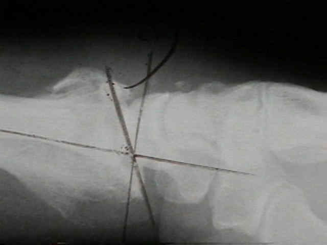
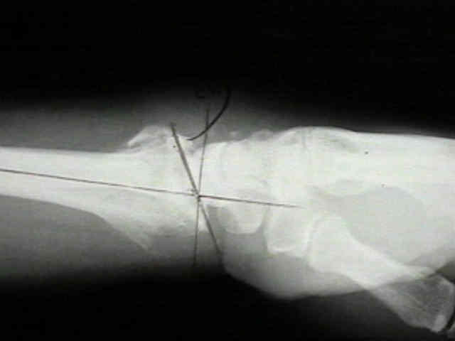
- Intrafocal (Kapandji) pinning of distal radius fractures with and without external fixation.
- ulnar styloid fracture:
- Radiographic Healing and Functional Outcomes of Untreated Ulnar Styloid Fractures Following Volar Plate Fixation of Distal Radius Fractures: A Prospective Analysis.
- rotational alignment:
- in the study by Tornetta, et al (15th Annual Meeting of the Orthopaedic Trauma Association, 1999), the authors point out
that there can be up to 38 deg of mal-rotation can be present before a step off can be appreciated on the lateral view;
- PA views were even less sensitive for determining malrotation;
- it was determined that over pronation of the distal fragment was associated with a more volar position of the ulna on a
true lateral view (where as it is normally slightly dorsal on a true lateral view);
- classification:
- Frykman Classification
- Melone Classification
- Universal Classification
- Routine Views:
- PA View
- radial inclination
- radial length: (ulnar variance)
- radial ulnar joints:
- distal radioulnar joint should measure approximately 2 mm;
- if there is a of a radio-ulnar joint disruption consider CT scan;
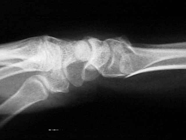
- lateral view
- fat pads: (in the case of occult injury)
- dorsal tilt:
- look for dorsal tilt of the lunate (DISI deformity);
- misc: consider use of a 20-25 deg tilted lateral to better profile the radial articular surface;
- ref: Tilted lateral radiographs in the evaluation of intra-articular distal radius fractures.
- Specialized Studies:

- Ulnar Deviation PA View
- Pronated Olblique (STT joint)
- Ulnar Deviation Lateral View:
- normally ulnar deviation will cause the lunate to dorsiflex and shift volarly, and the radio-luno-
capitate alignment resembles a DISI pattern;
- volar shift of the lunate helps maintain the normal co-linear relationship of the radius and
the capitate;
- w/ mid carpal ulnar instability, the lunate will dorsiflex, but will not have normal palmar translation;
- hence, the longitudinal axis of the capitate lies above the axis of the radius;
- this "zig zag" deformity would be expected to cause symptoms following distal radial fractures even
if the loss of volar tilt was minimal;
- CT scan: can help assess the step off of intra-articular fractures and comminution
Fractures of the distal radius. Intermediate and end results in relation to radiologic parameters.
Computerized tomographic evaluation of acute distal radial fractures.
Colles fracture: does the anatomical result affect the final function?
Colles' fracture. How should its displacement be measured and how should it be immobilized?
Factors affecting the outcome of Colles' fracture: an anatomical and functional study.