

- Blood Supply to the Wrist
- Carpal Instability
- Differential Diagnosis
- Kienbock's disease
- Luno-Triquetral Coalition
- Perilunate Dislocations
- Scapholunate Instability
- Vascularity of the Lunate
- X-ray findings
- Discussion:
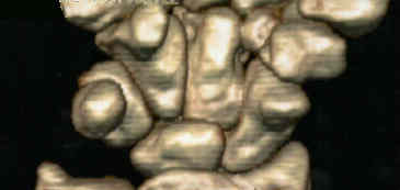
- lunate articulates proximally w/ radius and distally w/ capitate;
- it has large volar surface, & is displaced volarward w/ forced dorsiflexion of the wrist;
- most frequently dislocated carpal bone;
- w/ flexion capitate slides out from under lunate to create fullness where the capitate depression has been;
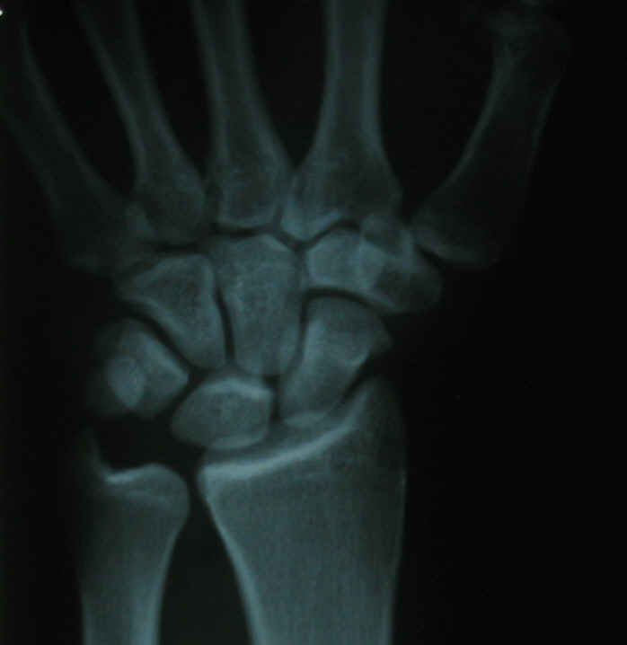
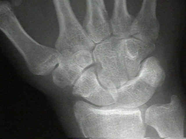
- Radiographs:
- lunate, capitate, and the base of the 3rd metacarpal are in line w/each other & is covered by base of ECRB;
- colinear alignment of: radius, lunate, capitate, & 3rd metacarpal;
- deviation of more than 15 deg either way between the links of chain may be viewed as lax, diseased, or damaged;
- Exam:
- it is palpable just distal to radial tubercle;
- w/ flexion and extension lunate/capitate articulation may be felt;
- tenderness of dorsal lunate may suggest Keinbock's dz, while more ulnar tenderness suggests tears of TFC or lunotriquetral ligament;
- knowing position of ECU & ulnar styloid helds to differentiate ECU tendinitis from distal radioulnar problems
Difficult wrist fractures. Perilunate fracture-dislocations of the wrist.