

- See: Radiology of the Elbow
- Discussion:
- note that accurate AP and Lateral views are essential to judge the reduction;
- slightly oblique views can lead to acceptance of malreduction;
- AP View:
- comparison films are usually required to judge amount of relative varus;
- generally no more than 4 deg of varus is accepted (as determined on by the Bauman's angle, seen on AP view):
- obtain AP radiograph of opposite extremity for comparison;
- note that inadequate radiographs may hide a "T" condylar fracture;
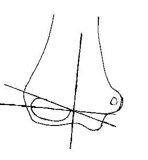
- Lateral View: 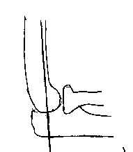
- insist on true lateral;
- normally the anterior humeral line should pass through ossification center of lateral condyle (this also holds true for type I frx);
- called the anterior humeral line;
- w/ type II frx:
- anterior humeral line does not transect capitellum;
- there should be a radiolucent interval between ossification center of capitellum and the semilunar notch of the olecranon;
- any overlap of the capitulum and semilunar notch implies malrotation;
- condylar - humeral shaft angle:
- needs to be restored in order to restore full flexion;
- soft tissue structures:
- superficial part of anterior fat pad should be in front of coronoid fossa;
- in normal elbow the anterior fat pad should be barely visualized & posterior fat pad should not be seen at all;
- look for small radiolucent area between bony rim & moderate opaque shadows of brachialis;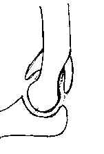
- w/ effusion 2nd to type I frx, there will be anterior & superior displacement of anterior fat pad;
- Oblique View:
- may be useful for diagnosing type I extension frx
Supracondylar fractures of the humerus. Assessment of cubitus varus by the Baumann angle.
Determination of medial epicondylar epiphyseal angle for supracondylar humeral fractures in children.
The posterior fat pad sign in association with occult fracture of the elbow in children.