

- See:
- Baumann's Angle
- Cubitus Varus
- Discussion:
- in general a varus reduction cannot be accepted whereas a slight valgus reduction can be accepted;
- the true amount of varus cannot be determined until arm is fully extended which is not performed until child is under anesthesia;
- Technique of Reduction:
- note that attempts in the ER at partial reduction and delays in reduction will only lead to increase soft tissue swelling which will
complicate the definative redection under GEA in the OR;
- arm is positioned on a sterile flouroscope which serves as a platform;
- alternatively the arm can be placed on a narrow hand table, so that the flouro table is free to rotate w/o having to rotate the arm;
- consider placing sheet around child's body to provide counter-traction during the reduction;
- first achieve adequate reduction prior to pin insertion;
- technique of percutaneous pinning is facilitated by a near anatomic reduction, and in contrast, a poor reduction will only
further complicate proper insertion of pins;
- the teaching point is that pins should not be inserted until an acceptable reduction is obtained;
- position the flouroscopy monitor, so the operator can look straight ahead;
- closed reduction relies on an intact periosteal hinge;
- shaft condylar angle needs to be restored inorder to avoid loss of flexion;
- rotational alignment needs to be restored inorder to avoid cubitus varus;
- beaware of interposed tissues:
- brachialis may become interposed at frx site or by proximal humeral frx fragment button-holed thru brachialis;
- if this occurs, closed reduction w/ longitudinal traction may worsen tightening of the muscle around protruding fragment;
- if the brachialis is buttonholed by the distal humeral spike, then the muscle can be milked off the spike by grasping the
proximal arm and squeezing sequentially from proximal to distal;
- avoid excessive medial squeezing (to avoid N/V injury);
- periosteum:
- tears proximal to frx site, and may become interposed into anterior frx gap;
- w/ interposed periosteum open reduction is required;
- First Restore Length:
- first restore length, followed by realignment of distal frag to humeral shaft;
- traction is applied to the arm to free the proximal fracture site from the brachialis muscle;
- then apply longitudinal traction w/ elbow in extension and supination in attempt to oppose edges of both fragments;
- counter traction is provided by the assistant;
- Medial and Lateral Translation and Rotation:
- fracture displacement indicates whether the medial or lateral periosteum is intact;
- while traction is being maintained, next the medial or lateral displacement of distal frag is corrected;
- during reduction, a small amount of medial or lateral displacement or small amount of anterior or posterior angulation may be
tolerated, but any malrotation is not acceptable;
- reduction is facilitated either by pronation (common) or supination (uncommon) depending on whether there is medial or
lateral frx displacement;
- medial displacement of distal fragment;
- more comon than lateral displacment;
- implies that the medial hinge is intact;
- this fragment needs to be laterally translated for reduction;
- ponation places the medial periosteum on tension, which closes the hinge and correcting varus malalignment;
- forearm is pronated to tighten the medial periosteal hinge along w/ application of valgus force and translation w/ one hand;
- traction must be maintained during this maneuver;
- full pronation is required to reduce the varus tilt;
- lateral displacement of distal fragment;
- forearm is supinated to tighten the lateral periosteal hinge along w/ application of varus force and translation w/ one hand;
- traction must be maintained during this maneuver;
- this tenant has been recently challenged by Khare, et al (1991) who noted that forearm pronation was more effective than
supination in reducing posterolaterally displaced supracondylar frx;
- references: Prevention of cubitus varus deformity in supracondylar fractures of the humerus.
- at this point, frx is out to length, and rotation has been restored;
- ensure that carrying angle is adequate (compared to opposite);
- note that if the internal rotation deformity is not corrected, then the varus deformity will not be corrected;
- Frx Angulation:
- next, address frx angulation and displacement;
- w/ extension type fractures, the posterior periosteum is intact which is used to facilitate the reduction;
- elbow is flexed to 120 deg while pushing distal fragment anterior to reduce it;
- this can be effected by placing thumb over olecranon and by pronating the forearm as the elbow is being flexed;
- similarly, a posterior directed force is applied to the proximal fragment;
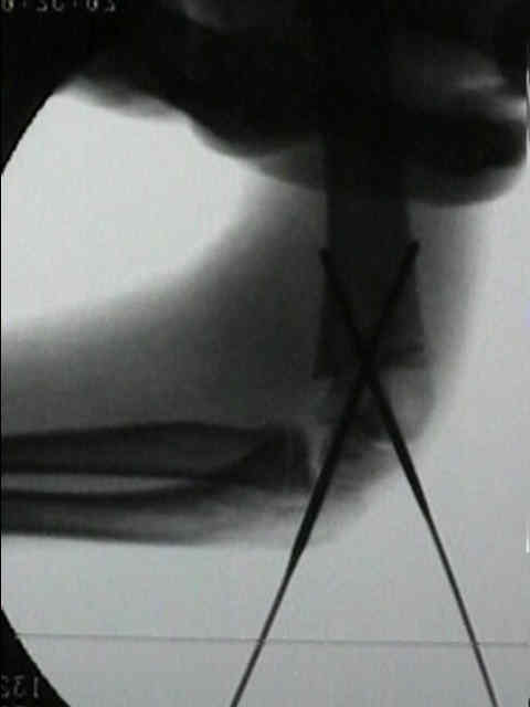
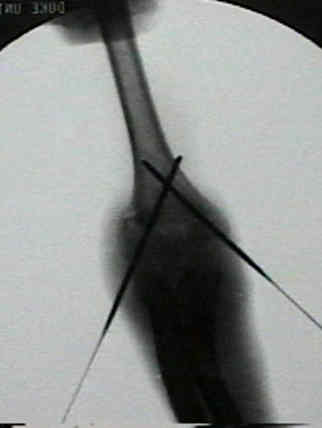
- frx stabilization:
- flexion is maintained to hold reduction while crossed pins are inserted;
- w/ anatomic reduction full flexion should be achieved;
- incomplete flexion, implies interposed soft tissue;
- Radiographs:
- comparison films are usually required to judge amount of relative varus;
- generally no more than 4 deg of varus is accepted (as determined on by the Bauman's angle, seen on AP view):
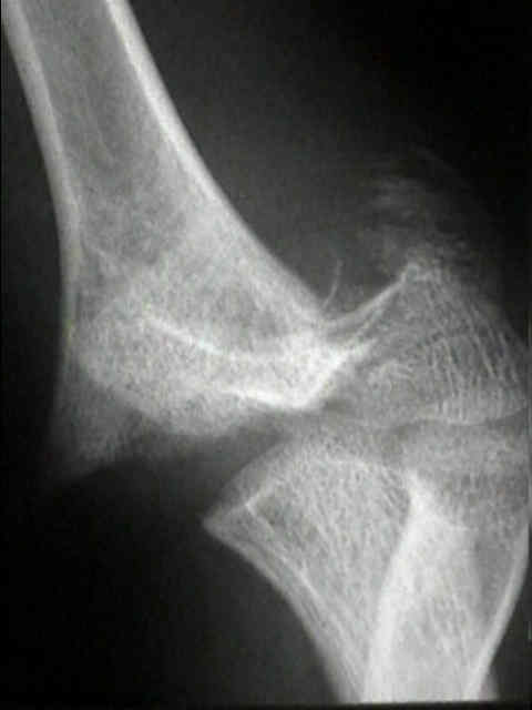
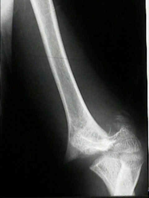
- on lateral view, anterior humeral line normally should pass thru ossification center of the lateral condyle (this also holds
true for type I frx);
- on lateral view, should be a radiolucent interval between ossification center of capitellum and semilunar notch of olecranon;
- any overlap of the capitulum and semilunar notch implies malrotation;
- the presence of an oblique spike indicates malrotation and the need for re-reduction;
- condylar - humeral shaft angle:
- needs to be restored inorder to restore full flexion;
- CT Scan:
- one unique application of CT imaging has been its use in determining adequacy of the reduction of supracondylar fractures
Clinical significance of anterior humeral line in supracondylar humeral fractures in children.