

- Anatomy of Olecranon: 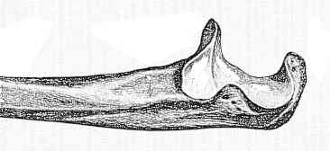
- Radiology:
- insist on a true lateral X-ray of the elbow joint to evaluate:
- extent of frx & displacement:
- non comminuted = displacement of less than 2 mm;
- w/ flexion, frx displacement may be found to increase;
- usually frx gaps dorsally, and some separation of fragments occurs, which leads to joint incongruity;
- degree of comminution;
- disruption of articlar surface in semilunar notch;
- displacement of radial head
- definition of displacement:
- displacement of > 2 mm;
- increase in degree of separation w/ 90 deg. flexion of the elbow;
- inability to extend the elbow actively against gravity
- Mayo classification
- type I: (undisplaced fracture);
- by definition is stable to flexion and extension and allows early mobilization in 3-5 days;
- type II olecranon frx:
- displaced, stable frx;
- frx fragments are displaced > 2-3 mm, but collateral ligaments are intact;]
- frx may be non-comminuted (Type IIA) or comminuted (Type IIB).
- no sign of subluxation;
- type III:
- displaced fracture:
- frx is displaced & forearm is unstable in relation to humerus;
- this injury is really a fracture-dislocation.
- misc:
- coronoid process + olecranon frx;
- patella buitis:
- ocassionaly occurance of patella buitis, which is true accessory ossicle located in triceps tendon at its insertion into olecranon;
- may be confused with a fracture
Some vagaries of the olecranon.
Olecranon stress fractures in throwers. A report of two cases and a review of the literature.