 - See:
- Extensor Mechanism Injuries of the Knee
- Quadriceps
- Patellar Tendon Avulsion from TKR
- Discussion:
- usually occur in pts under age of 40;
- most ruptures occur w/ the knee in a flexed position (around 60 deg) which are then subject to excessive loading;
- great majority occur at level of inferior patellar pole (> level of tibial tubercle);
- patellar tendon avulsions occur more often in person of african decent (? greater proportion of quadriceps type II muslce fibers);
- Exam:
- need to distinguish partial rupture from complete rupture;
- reveals palpable defect in patellar ligament;
- high riding patella;
- inability to extend knee (complete tears);
- partial tears are characterized by ability to extend knee, but full extension may be lacking;
- note patients w/ partial patellar tendon ruptures may not be able to extend the knee due to pain and knee effusion;
- consider knee aspiration and injection of lidocaine prior to asking the patient to extend the knee;

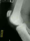
- Lateral Radiograph:
- may reveal small avulsion from inferior patellar pole
- significant patella alta is evident using Insall method, or using the Blackburne method;
- inferior patellar border will lie above Blumensaat's line;
- Non Operative Treatment:
- only for partial patellar ligament disruption;
- partial tears can be treated w/ immobilization for 3-6 weeks;
- complete tears of the patellar ligament require surgical repair;
- end-to-end suturing may not be attainable for delayed or late repair;
- Operative Technique: using end-to-end sutures;
- equipment:
- power drill for 2.5 mm drill bit;
- wire passer or 25 gauge wire and 14 gauge angiocath (for passing sutures through the patellar drill holes);
- No 5 Ethibond (or equivalent) and appropriately sized tapered free needles;
- patient is positioned supine with a bump under the hip;
- make longitudinal incision from inferior pole of patella to the tibial tubercle;
- identify the adjacent retinacula on either side of the tendon;
- if the retinaculum is torn, it can be helpful to place clamped-untied sutures early on, rather than at the end of the case;
- frayed tendon edges are sharply debrided;
- horizontal trough is made in the inferior patellar surface inorder to promote healing;
- tendon stitch statedgy:
- generally two double armed No 5 sutures are passed from distal to proximal so that 4 suture strands pass out of the free tendon edge;
- augmented becker technique:
- in the study by Singer, et al (1998), the core suture technique was the most important element in establishing both strength and stiffness of the repair;
- the Becker technique involves 4 strand repair with two knots out side of the repair site;
- repair consists of criss-crossing running suture using a double armed needle;
- sutures should be placed 0.75 cm from the cut edge of the tendon;
- volar epitenon suture is used to augment the repair;
- as noted in the report by Howard, et al (1997), the MGH tendon repair technique (crossing running suture repair) was signficantly more resistant to gap formation than the Bunnel or the Krackow technique;
- MGH tendon repair has superior suture purchase which is probably related to superior resistance to gap formation;
- Use of the Taguchi method for biomechanical comparison of flexor-tendon-repair techniques to allow immediate active flexion. A new method of analysis and optimization of technique to improve the quality of the repair.
- Biomechanical analysis of four-strand extensor tendon repair techniques.
- patellar drill holes:
- w/ this statedgy, three vertically oriented drill holes are made in the patella, which allow both suture arms to be carried thru patella and tied over top (from Lindy, et al (1995)).
- at the inferior pole of the patella, these drill holes must exit near the articular surface to prevent abnormal tilting of the patella w/ flexion;
- create 3 evenly spaced vertically oriented drill holes thru the patella;
- sutures are passed thru the drill holes (from distal to proximal) using a Huston wire passer;
- if a Hughston wire passer is not available, pass an angiocatheter through the drill hole (distal to proximal), and then pass a thin wire into angiocatheter (proximal to distal);
- once the wire is passed through the drill hole, make a small loop on the distal end, which allows sutures to be pulled thru the drill holes (from distal to proximal);
- the outer suture limbs are carried thru the outer holes, and both central suture limbs are carried thru the middle hole, and are then tied;
- Repair of Patellar Tendon Disruptions without Hardware.
- judgement of patellar height:
- before sutures are tied down, check patellar height;
- at 45 deg of knee flexion, the inferior pole of the patella should lie just above the notch;
- residual gaps in the tendon repair are repaired w/ a running 2-0 Viccryl Suture;
- retinacular repair:
- if the repair is strong, consider not repairing the lateral retinaculum (if it is torn) inorder to avoid patellar subluxation;
- re-enforcement technique:
- serves as an adjunt to suture tendon repair and protects the tendon repair as the patient is started on early motion;
- turndown technique:
- inverted V can be created out of the patellar periosteum which is then turned down in order to augment the repair;
- suprapatellar augmentation:
- 5 mm Mersilene tape is passed thru a transverse drill hole made through the tibial tubercle, is then woven thru the para-patellar retinaculum on both sides;
- second strand of tape is then passed through the middle of the quadriceps insertion on patella;
- tape is tied on both sides w/ the knee in 90 deg of flexion (after tourniquet is released);
- tightening of the Mersilene tape w/ the knee in extension or tightening to the point that the patella is brought distally will often result in patellar infera and patellofemoral chondrosis (Lindy, et al (1995));
- some patients will complain of prominent tape knots, which can be later removed;
- Repair of Patellar Tendon Disruptions without Hardware.
- use of wire for augmentation:
- while use of 18 gauge wire is effective for augmentation, it often causes causes local tenderness;
- the patient should be forwarned that the wires frequently break and will typically require removal;
- Post Op ROM:
- historically, knee was immobilized in extension for 6 weeks, however, many surgeons are now permitting passive ROM in the immediate postop phase;
- full wt bearing is allowed (using knee immobilizer)
Late reconstruction of the patellar tendon.
Repair of Patellar Tendon Disruptions without Hardware.
A systematic approach to reconstruction of neglected tears of the patellar tendon. A case report.

