

- See: Patella Alta:
- Discussion:
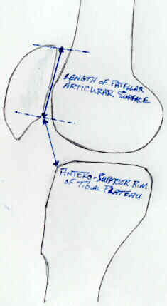
- patellar height in relation to the joint line: (2 methods)
- Insall and Salvati Method
- Blackburne and Peel Method
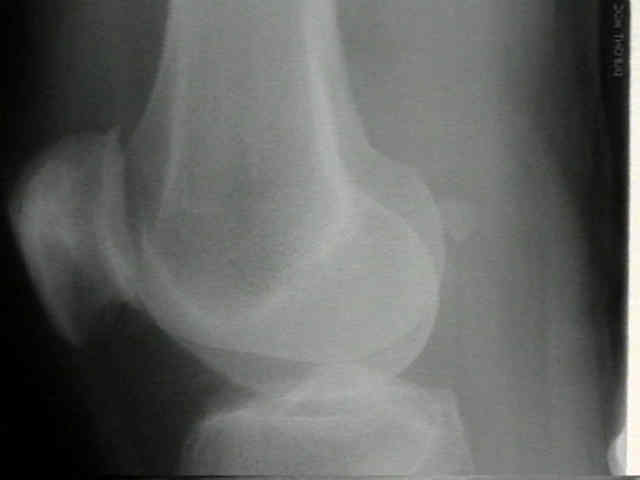
- fat density zone seen posterior to the quadriceps (water density) represents suprapatellar fat pad;
- fat pad will be displaced anteriorly with joint effusion;
- fabella:
- sesamoid bone embedded in lateral head of gastroc muscle;
- seen in about 10% to 20% of individuals;
- technique:
- pt lies flat on the affected side with the knee flexed 25-30 deg;
- central beam is directed toward the medial aspect of knee joint w/ 5-7 degrees of cephalic tilt