- See:

- FDP Lacerations:
- Rupture of FDP
- Forearm Flexors
- Quadriga
- Anatomy:
- origin:
- upper 3/4 of anterior & medial surfaces of ulna, interosseous membrane and deep fascia of the forearm;
- FDP is dorsal to FDS in the volar aspect of the forearm
- insertion:
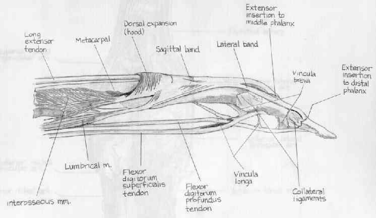
- four tendons (one to each finger) to palmer base of distal phalanx, after passing thru tendon of FDS;
- distal anatomy:
- lumbrical muscles arise from the FDP tendons and course distally to their insertion into the radial lateral bands of the digits;
- at the entrance to the fibro-osseous tunnel (A1 pulley), FDS bifurcates, permitting FDS to course superficial onto its insertion;
- synergists: FDS;
- nerve supply:
- AIN branch ofmedian nerve supplies index & middle fingers in 75% of cases: C8 > C7, T1
- ulnar nerve supplies middle, ring & little fingers in 75% of cases (therefore middle finger has dual innervation in 75% of patients): C8 > C7, T1;
- ref: Variations in innervationn of the flexor digitorum profundus muslce.
- anatomy discussion:
- FDP originates high on the on the forearm & divides into 4 parts;
- segment to index finger divides proximally & is partially separated from tendons directed to the ulnar 3 digits, which are closely interelated in the midpalmar area;
- ulnar 3 digits share common muscle belly, and thus independent flexion of any finger w/ others restrained in extension requires intact FDS functions to that finger;
- tendons of the profundus do not divide until they reach the wrist;
- this enables the superficialis tendons to act more independently but affords greater power to profundus tendons;
- individual flexion of index finger is possible to about 30 deg, at point of about 30 deg other profundi begin to f(x);
- FDP tendons at wrist are covered by synovial sheath that is thickened by aging, RA, or trauma;
- enlargement of this sheath fills carpal canal & results in compression of median nerve;
- tendons of the profundus lie side by side at wrist, whereas tendons of superficialis are arranged so that those for 3rd & 4th digits are
anterior to those of the second and fifth digits;
- FDP tendon is attached to FDS by vincula, & both are encapsulated by annular ligament and a synovial sheath;
- FDP tendon inserts into distal phalanx after passing over volar capsule of the distal joint;
- Exam for FDP Injury:
- FDP tendons of ulnar 3 digits have minimal individualization;
- if one digit is held in extension, only limited flexion of adjacent DIP joints is possible;
- if index finger is held in extension, ulnar 3 digits can be flexed almost to palm;
- isolated testing of the FDP is performed by having middle phalanx held in full extension by the examiner;
- distal phalanx is then actively flexed;
- test is best performed by maximally extending wrist and MP joints;
- ie, examiner's holding all finger joints in extension except distal joint;
- FDP to index finger shows isolated function in 85 % of pts;
- in 15% there is simultaneous funciton w/ FPL
- in FDP, ulnar 3 digits act as single unit & are tested simultaneously;
- FDP tendons are connected to each other by multiple bands of tendons, which results in simultaneous action of 3rd, 4th, & 5th fingers;
- if one of these fingers is held in flexion, the other 2 cannot be straightened completely or, if one digit is kept in extension, flexion of
adjacent fingers cannot be done individually;
- lesser number of bands extend from ulnar 3 digits to index finger;
- for this reason, the index finger functions more independently;
- thumb functions most independently, but in 10 % of general population tip of thumb and index finger f(x) simultaneously
- FDP Lacerations:
Results of transfer of the flexor digitorum superficialis tendons to the flexor digitorum profundus tendons in adults with acquired spasticity of the hand.
Two-stage flexor-tendon reconstruction. Ten-year experience.
Anatomy of the flexor retinaculum.
Avulsion of the profundus tendon insertion in athletes.
Anomalous tendon slips from the flexor pollicis longus to the flexor digitorum profundus.
Variations in innervationn of the flexor digitorum profundus muscle.


