

- See: TKR: Soft Tissue Balance and Collateral Ligaments
- Discussion:
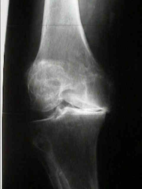
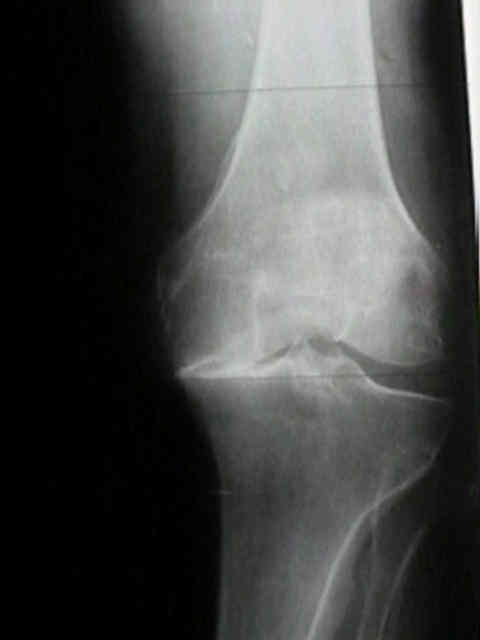
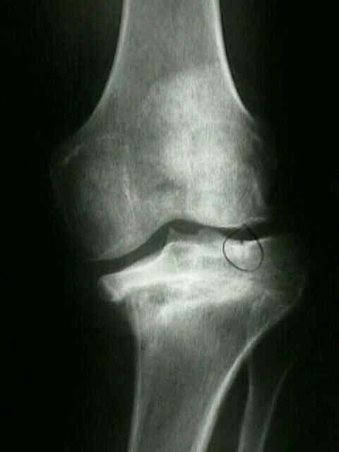
- fixed varus is the most common angular contracture in pts undergoing tkr;
- this deformity may be accentuated by the presence of medial tibial plateau or medial femoral condyle osteophytes;
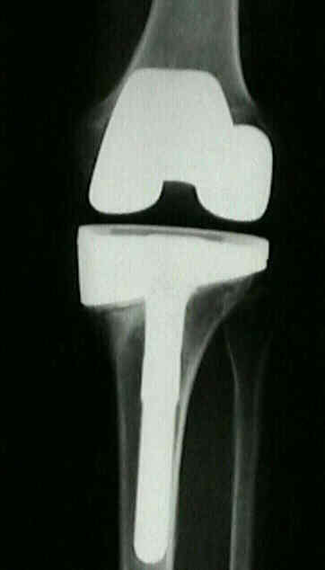
- it is essential that the varus alignment be corrected at the time of surgery;
- failure to correct varus alignment will signficantly increase contact stresses on the polyethylene comonent;
- unconstrained designs which have a flat configuration on the femoral and tibial surfaces in the coronal plane (such as the
Advantium (Wright medical), Miller Galante II (Zimmer), and Omnifit (Osteonics) will show increase in polyethylene
contact stresses if tibial component is placed in varus alignment;
- in contrast, components that have tibial and femoral components which are matched in the coronal plane are more tolerant of varus
alignment of the tibial component (examples are the Profix (Richards) and LCS (Depuy);
- effects of tibial varus deformity on posterior condylar angle: (femoral component rotation);
- in 60 consecutive total knee arthroplasties done in 52 patients with primary osteoarthritis and varus or neutral tibiofemoral
alignment, posterior condylar angle was calculated intraoperatively and averaged 3.98° (range, 0°-9°);
- 18 knees had a posterior condylar angle value less than 3° whereas 27 knees had a posterior condylar angle value of 5° or greater;
- final rotational alignment of the femoral component was set parallel to the transepicondylar axis;
- only one of these 60 knees required a lateral retinacular release for proper patellar tracking during the knee arthroplasty;
- when compared with three previously defined angles measured on the radiographs taken preoperatively, only the tibial
plateau tibial shaft angle values were correlated significantly with the value of the posterior condylar angle;
- as tibial varus joint line obliquity increased, there is tendency for the transepicondylar axis to be rotated more externally
relative to posterior condylar axis;
- this variance suggests that the use of the posterior condylar axis as a rotational reference is inappropriate in many knees
w/ arthritis w/ varus or neutral tibiofemoral alignment;
- in particular, varus tibial joint line obliquity of more than 4 deg increases the likelihood of femoral component
malrotation when posterior femoral condyles are used to reference femoral component rotation;
- ref: Varus Tibial Joint Line Obliquity: a Potential Cause of Femoral Component Malrotation.
- varus tibial component positioning has lead to increased failure, radiolucent lines, and medial compression
- 12/14 TKAs with tibia in neutral or varus failed (Bargren, et al (1983))
- varus-cut tibia has statistically highest avg peak strain in posteromedial tibial plateau
- hot spot on all varus aligned tibia and none of the neutral tibia
- varus alignment may cause medial compartment overload, cancellous bone collapse, and failure of TKA
- neutral alignment has protective effect against medial loading
- may explain an early failure mechanism in malaligned TKA
- altering tibiofemoral alignment by cutting the femur in more valgus does not compensate for this error
- references:
- Alignment in total knee arthroplasty. Correlated biomechanical and clinical observations.
- The effects of varus tibial alignment on proximal tibial surface strain in total knee arthroplasty: The posteromedial hot spot.
- Preoperative Planning::
- May obtain full-length lower extremity AP/Lat x-ray; evaluate anatomic axis:, mechanical axis:
- esp. unusual cases: excessive femoral bowing or malunion, reduced hip offset
- also helps to obtain desired entry hole position in femur for intramedullary guide
- films can be misleading, must be in neutral rotation
- ref: Sequential release for soft tissue balancing during TKAs for varus knees after femoral and tibial cuts:
- cautions:
- note whether varus/valgus deformities are fixed vs flexible;
- it is important to distinguish between a fixed varus knee and a knee w/ pseudolaxity due to loss of the medial joint space;
- in the later case, the MCL may be attenuated and can easily be "overstripped" during the initial exposure (when this happens, a
larger spacer is needed to restore stability);
- w/ a fixed varus knee, further capsular elevation may be required;
- Yagishita et al (2003) suggest
1- removal of ACL, PCL and menisci
- w/ residual varus contracture, PCL is released, which of course, requires insertion of posterior stabilized knee;
- although PCL release is not necessary when performing a TKR in non contracted knee, its required in cases of severe fixed
varus, since it is a deforming force;
2- subperiosteal release of deep MCL fibers
3- release of medial and posteromedial capsule
4- resection of femoral/tibial medial osteophytes
5- release of tibial insertion of superficial MCL (8-10 cm distal to medial joint line)
- Reference: Step-by-step measurements of soft tissue balancing during total knee arthroplasty for patients with varus knees
- Insall et al (1985) suggest
1- Remove femoral and tibial peripheral and posterior osteophytes and alignment is reassessed
2- Remove menisci with its capsular attachment
3- Subperiosteal release of deep MCL attachment to tibia with initial approach (see MCL)
- if neutral alignment is not achieved, capsular sleeve is dissected subperiosteally beyond the midcoronal plane to level
of posteromedial corner of the joint;
- be careful not to strip the posterior oblique ligament;
- in most cases, correction will be achieved w/ reflection of deep MCL and only rarely is reflection of the complete insertion
of superficial MCL required;
- medial capsular sleeve can be released (or recessed) distally and allowed to slide proximally;
- incise the periosteum of the tibia along the anterior crest, extending approximately 2-4 cm below joint line;
- continue the reflection of the capsule distally and posteriorly, elevating periosteum, coronary ligaments, pes anserinus,
MCL, and capsule in one continuous layer;
- valgus instability is avoided by properly balancing the flexion and extension spaces with a thick enough polyethylene
implant inorder to tense the medial capsular sleeve;
4- Subperiosteal release of distal insertion of superficial MCL
- superficial MCL is associated intimately w/ pes anserinus, even complete elevation from tibia does not cause gross
instability, as can occur w/ complete release from femur;
- popliteus contracture:
- with many severe fixed varus deformities there will be associated lateral subluxation of the tibia on femur (varus thrus;
- this is the result of contracture of popliteus & is corrected by tenotomy of popliteus tendon at time of fixed varus release;
- releasing popliteus tendon may also improve knee flexion;
- if the popliteus is left intact, it should be rechecked once the trial components are in place;
- in this case, the tendon may be tight, because of rotational alignment of femoral component;
- trial components and preparation for revision components:
- with proper ligament balancing, there still may be unacceptable laxity or instability;
- in this case, revision constrained components may be required
- Case Example:
The effect of extraarticular varus and valgus deformity on total knee arthroplasty.
The effect of varus tilt on contact stresses in total knee arthroplasty: a biomechanical study.
The Difficult Knee: Severe Varus and Valgus.
Efficacy and Safety of a Novel Three-Step Medial Release Technique in Varus Total Knee Arthroplasty
Postoperative Lateral Ligamentous Laxity Diminishes with Time After TKA in the Varus Knee