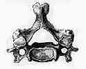
- Discussion:
- characterized by weakness (upper > lower extremity);
- ataxic broad based suffling gait, sensory changes;
- rarely urinary retention;
- anatomy of compression:
- anterior cord compression ---- protruding disc or posterior osteophytes;
- anterolateral compression ---- joints of Luschka
- lateral compression ---- cervical facets
- posterior compression ---- ligamentum flavum
- differential diagnosis:
- multiple sclerosis
- unlike cervical myeopathy there is abnormal cranial nerves, and jaw jerk;
- amyotrophic lateral sclerosis
- affects both upper and lower motor neurons, and no alterations in sensation;
- SCIWORA syndrome
- spinal cord tumor
- syringomyelia
- subacute combined degeneration
- in C.M. there is more sensory loss in the upper extremities and more spastic lower extremities;
- cerebral hemisphere lesion
- low pressure hydrocephalus
- catagories:
- transverse lesion syndrome:
- corticospinal, spinothalamic, and posterior cord tracts are involved equally;
- associated with the longest duration of symptoms
- may represent end stage of the disease
- motor system syndrome:
- corticospinal tracts and anterior horn cells are injured causing spasticity;
- central cord syndrome:
- motor and sensory deficits affected the upper extremities more severely than the lower extremities;
- Brown-Séquard syndrome:
- ipsilateral motor deficits with contralateral sensory deficits
- may be the least advanced form of the disease;
- Brachialgia and cord syndrome: radicular pain in the upper extremity along with motor and/or sensory long-tract signs.
- Clinical Findings:
- see: exam of spinal cord
- presentation: (Bertalanffy, et al)
- 61% presented with radicular symptoms
- 16% had pure myelopathic symptoms
- 23% had a combination of myelopathic and radiculopathy
- upper motor neuron findings such as hyper-reflexia, clonus, or Babinski's sign may be present;
- funicular pain, characterized by central burning and stinging w/ or w/o (Lhermitte's phenomenon - radiatineg lightening like sensations down back w/ neck
flexion) may also be present w/ myelopathy;
- ref: Complications of anterior cervical discectomy without fusion in 450 consecutive patients.
- jaw jerk:
- the jaw jerk, which is controlled by the fifth cranial nerve is performed by the tapping on the slightly opened jaw;
- a normal reflex contraction of the masseter effectively rules out pathology above the foramen magnum;
- upper extremity: - mixed upper and lower motor neuron findings;
- myelopathy can initially present w/ hand dysfunction w/ loss of fine motor function such as writing;
- Reflex Testing:
- Hoffman's Sign:
- myelopathic hand:
- finger escape sign (small finger spontaneously abducts due to weak intrinsics) indicating cervical myelopathy;
- inverted radial reflex: (C5 - C6)
- inverted radial reflex may be present when cord & root compression are present at the C5 level;
- this reflex is demonstrated by tapping brachioradialis tendon;
- diminished reflex is noted along with a reflex contraction of spastic finger flexors;
- this specific reflex occurs due to peripheral compression of the C6 nerve root (from disc or spur) which allows upper motor neuron reflex to occur;
- biceps reflex primarily indicates neurologic integrity of C5;
- the reflex also has a C6 component;
- lower extremity - upper motor neuron signs;
- lateral cord involvement: causes spasticity, hyper-reflexia, and frank clonus in lower extremities;
- posterior cord involvement: causes decline in ability to walk, apparent ataxia;
- loss of lower extremity proprioception;
- Lhermitte's sign:
- paresthesias or leg weakness exacerbated by neck flexion;
- men may be cursed by this as they attempt to urinate;
- ref: Lhermitte's sign: Review with special emphasis in oncology practice.
- Babinski's sign:
- may not be present until myelopathy becomes severe;
- upper motor neuron findings such as hyper-reflexia, clonus, or Babinski's sign may be present;
- ref: Prevalence of Physical Signs in Cervical Myelopathy: A Prospective, Controlled Study.
- EMG:
- Radiographs:
- flexion and extension views:
- lateral view:
- consider:
- developmetal stenosis:
- canal diameter to body diameter should be greater than 0.8;
- spondylosis
- cervical kyphosis:
- old dens frx non union
- MRI:
- w/ subtle clinical and x-ray findings consider dynamic MRI (flexion-extension)
- spinal cord may show increased signal changes on T2 images;
- CT Myelogram:
- compression ratio: smallest AP diameter divided by largest transverse diameter;
- Treatment
- prognosis:
- condition has high potential for becoming worse leading to severe disability;
- myelopathic symptoms have variable potential for recovery, but prognosis for recovery is better when decompression is performed early;
- Anterior Arthrodesis and Decompression
Upper-airway obstruction after multilevel cervical corpectomy for myelopathy.
The surgical management of cervical spondylotic radiculopathy and myelopathy.
Subtotal vertebrectomy and spinal fusion for cervical spondylolytic myelopathy.
Anterior surgery for cervical spondylotic myelopathy: Smith-Robinson, Cloward, and vertebrectomy.

