

- See: Myelodysplasia:
- Discussion:
- syringomyelia is a condition in which tubular cavity, or syrinx, in central area of spinal cord gradually expands leading to progressive myelopathy;
- it is associated w/ disturbed hydrodynamics of cervical spinal fluid w/ or w/o associated obstruction at the cranial cervical junction;
- syrinx develops most often in the cervial and thoracic regions;
- it may be idiopathic (occuring during first few decades of life) or may be aquired;
- almost all pts w/ syringomyelia have type I Arnold Chiari malformation, which may produce symptoms of medullary or upper cervical compression;
- diff dx: central cord syndrome:
- causative conditions:
- spinal cord injury:
- may occur in 1-3% of patients w/ spinal cord injury;
- sometimes the syrinx may become apparent only years after the injury;
- syrinxes develop twice as often in the thoracic spine as the C-spine;
- syrinxes tend to occur at the level of the original trauma, but often will extend cephalad from the level of the spinal injury;
- occur more often in patients w/ complete spinal cord injury or in patients w/ GSW to the spine;
- spinal cord neoplasia:
- associated conditions:
- scoliosis:
- look for rapid progression of curve;
- left thoracic scoliosis is common;
- treatment of the scoliosis without recognition of syringomyelia and Chiari malformation can lead to paraplegia;
- Klippel-Feil syndrome;
- Arnold Chiari malformation
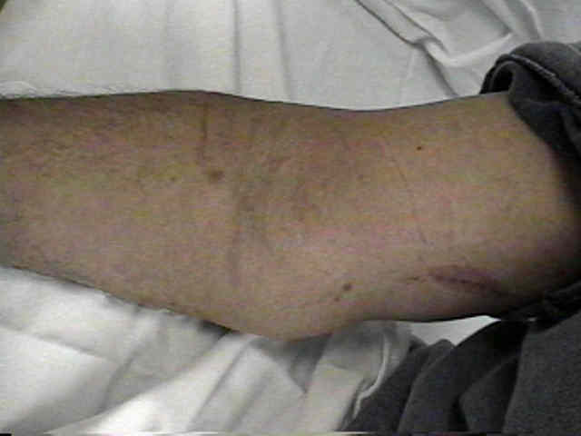
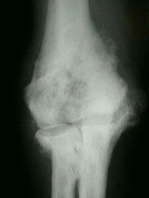
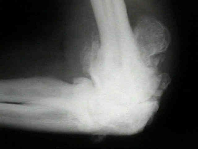
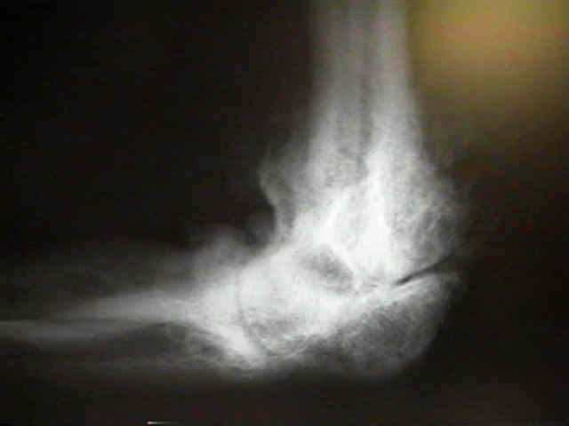
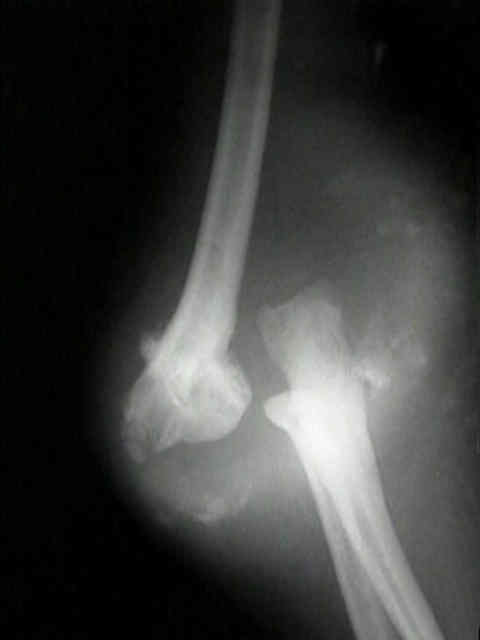
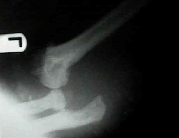
- neuropathic joints:
- neuropathic shoulder:
- most commonly involved joint;
- neuropathic elbow:
- look for associated ulnar and radial nerve entrapment at the elbow;
- ref: Neuropathic Arthropathy of the Elbow. A Report of Five Cases.
 ***
*** 
- Pathoanatomy:
- syrinx affects: long tracts, pyramidal tracts, & posterior column;
- serial sections show that it arises as a diverticulum from central canal of the spinal cord;
- anterior horn neurons crossing in central gray matter (spinothalamic fibers) are disrupted in early stages;
- clinical result is disruption of pain & temperature fibers at level of syrinx;
- pain and temperature sensation is preserved below the syrinx, because the spinothalamic tract is not affected by this central cord syndrome;
- touch and proprioception posterior column are affected in more advanced cases, due to more peripheral location of these nerve fibers;
- Clinical Findings:
- most common findings in post traumatic cases are radicular pain, spasticity, sensory loss, an weakness (often these symptoms are bilateral);
- valsalva maneuvers may worsen symptoms;
- lower motor neuron lesion is found at the level of dysfunction;
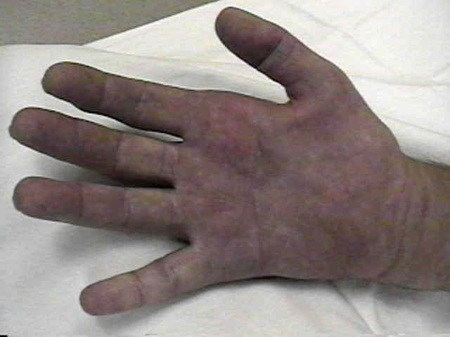
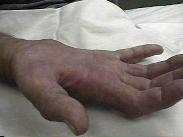
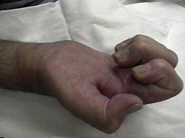
- look for atrophy and fasiculations of the hands and arms;
- scoliosis:
- scoliosis is associated with syringomyelia in 25% to 80% of cases;
- left thoracic scoliosis, rapid progression of curvature, or progressive neurologic symptoms is common in syringomyelia;
- MRI:
- on MRI, short TR images accurately show the morphology of the cyst;
- there is sharp interface between the normal cord & syrinx itself;
- metameric haustrations are typical of Chiari-associated hydromyelia;
- syrinx itself is often well defined and smooth, but occasionally septa are detected;
- if arachnoiditis is present, extensive adhesions may blur interface between cord & surrounding CSF;
- if cyst shows suspicious features at all, such as loss of definition in a focal region, then contrast enhancement is indicated to ensure that a
neoplasm is not present;
- note: that MRI may give distorted images if spinal hardware is present;
- in this case, consider CT myelogram;
- Treatment:
- w/ surgical drainage, patients can often expect improvement in radicular pain and sensory disturbances.
- motor function may return more often than sensory function;
- spasticity responds less predictably and often poorly to surgical drainage
Routine use of magnetic resonance imaging in idiopathic scoliosis patients less than eleven years of age.
Management of scoliosis due to syringomyelia in childhood and adolescence.
Progressive scoliosis as the first presenting sign of syringomyelia: Report of a case.
Orthopaedic features in the presentation of syringomyelia.
Syringomyelia and scoliosis. Huebert HT, MacKinnon WB: J Bone Joint Surg 1969;51B:338-343.
Syringomyelia and scoliosis in childhood and adolescence.
Management of scoliosis and syringomyelia in children.
Radiological Presentations in Relation to Curve Severity in Scoliosis Associated With Syringomyelia