

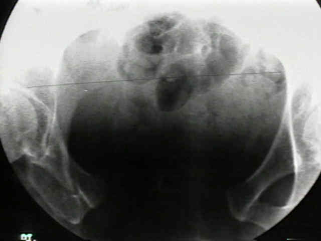
- Location of Pin Insertion Sites:
- depending on type of frame used, at least two or three pins are placed along iliac wing;
- 1st pin is usually placed about 2 cm posterior to anterosuperior spine.
- rest of pins should then be placed along iliac crest using external fixation clamp to determine the proper pin spacing;
- it may be difficult for straight external fixation clamp to a curved pelvis;
- because iliac crest overhangs on the lateral aspect, pins placed in this area tend to miss the iliac wing;
- Incision:
- closed technique is done thru percutaneous stab incisions 1-2 cm long at right angles to the iliac crest (transverse).
- these incisions allow the surgeon to find appropriate orientation of wing and, when reduction is accomplished, relieve excessive skin tension on pins;
- it is important to remember that the skin will tend to tent over the half pins following reduction of an open book injury;
- hence, make incision more medial than planned pin insertion site;
- also, attempt to reduce the fracture prior to pin insertion, which will also reduce soft tissue tension on the pins;
- open technique:
- an incision is made on either side of pelvis, from 1 cm distal to the ASIS extending proximally over the rim of the iliium;
- by means of a Cobb elevator place approximately 1 cm proximal to the ASIS, muscles are elevated medial and lateral to the iliac brim, in
order to allow palpation of the inner and outer tables of the pelvis during screw insertion;
- Pin Insertion Technique:
- location of iliac tables:
- spinal needles or small probes placed down inner & outer tables of pelvis help in the proper orientation of these pins;
- place pins in the outerhalf of the iliac crest, inorder to avoid perforating the cortex;
- pin direction:
- great care should be taken to avoid the tendency to drill in pins at right angles to the patient on the operating table, since this will cause the fixator to impinge on the thighs when the patient attempt to sit up;
- generally pins should be directed toward the acetabulum, so that the pins are angle toward the patients head;
- insert pins:
- then place a drill hole thru crest and then allow the pin to cut its own path between the iliac tables;
- if pins are directed in a caudal to cranial direction, there may be impingement on the pt's abdomen in the sitting position;
- palpate the ilium to assure that no inadvertent penetration of either cortex occurs as screw is inserted;
- pin placement can be confirmed at time of insertion by using flouro;
- Alternative Pins Sites:
- frontal pins: (between the ASIS and AIIS);
- strong pillar of bone runs from the AIIS to the posterior iliac crest and there is minimal soft tissue in this area;
- using an open technique, periosteal dissection is carried down over sartorius muscle & direct head of the rectus femoris;
- ASIS is identified, & pins are inserted about 1 cm apart between anterosuperior and anteroinferior spines.
- three pins can be used in this area & are best inserted w/ help of an image intensifier to avoid placement into the hip joint;
- insertion can be done percutaneously with the image intensifier:
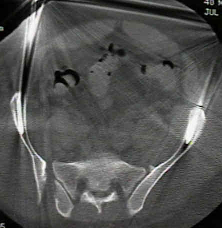
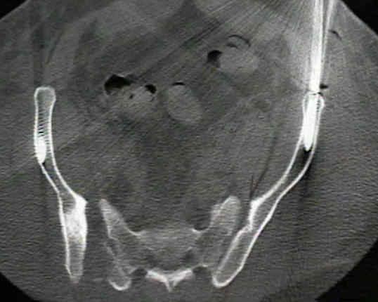
- obturator oblique view:
- a slight alternative view is the obturator outlet view which is a combination of the obturator view and the outlet view;
- this will show the safe zone (or corridor) as a tear drop;
- obturator oblique view shows the recommended half-pin insertion site approximately 2 cm above the hip (avoid hip joint)
- watch out for lateral femoral cutaneous nerve;
- iliac oblique view: shows the trajectory of pin insertion, to make sure the pin is passing superior to the superior gluteal notch;
- obturator inlet view: assures that the pin is contained within the bone for its entire length;
- hazards:
- pins may inadventently be directed into the hip joint;
- on occassion there is more bleeding than is expected;
- references:
- Placement of Half-Pins for Supra-acetabular External Fixation: an Anatomic Study.
- Supra-acetabular placement of external fixator pins: a safe and expedient method of providing the injured pelvis with stability.
- Anterior Pelvic External Fixation: Is There an Optimal Placement for the Supra-Acetabular Pin?
External fixation of unstable Malgaigne fractures: the comparative mechanical performance of a new configuration.
Anatomic and radiographic considerations in the placement of anterior pelvic external fixator pins.
Biomechanical testing of new and old fixation devices for vertical shear fractures of the pelvis.
Stabilization of unstable pelvic fractures with supraacetabular compression external fixation.
Effect of pin location on stability of pelvic external fixation.