

- Discussion:
- retrograde IM nailing
- Workup for Femoral Shaft Frx
- PreOp Planning:
- rule out femoral neck fracture:
- Is Reconstruction Nailing of All Femoral Shaft Fractures Cost Effective? A Decision Analysis.
- Diagnosis of femoral neck fracture associated with femoral shaft fracture: blinded comparison of computed tomography and plain radiography.
- IM nailing of pediatric femur frx
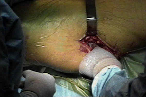
- Positioning:
- Frx Reduction:
- Proximal Frx;
- Distal Frx;
- Rotational Alignment:
- note rotational alignment of the opposite non injured side.
- a true lateral will define the plane of version;
- epicondylar axis is int rotated 20 deg (for 20 deg of anteversion)
- while obtaining true lateral, take time to mark out incision w/ ruler;
- acceptable reduction includes 8 deg of varus, 15 deg of valgus, malrotation of IR 15 deg and malrotation of ER 20 deg;
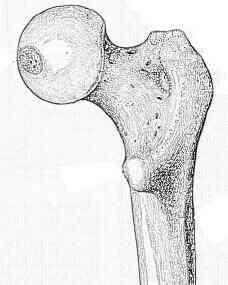
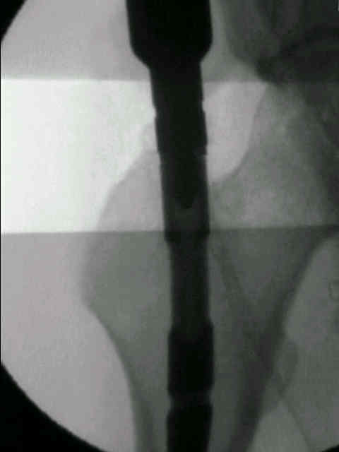
- Location of Canal Entry Point:
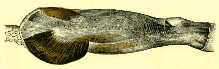
- hole is located just medial to greater troch in piriformis fossa;
- have assistant adduct proximal fragment (to better expose piriformis)
- use awl instead of steinman pin for initial entry:
- w/ steinman pin entry may be thwarted by a prominent trochanter;
- typically, must drop hand approx 20 deg and position awl up against the pts torso;
- then insert guide pin thru cannulated awl, and use awl to direct guide pin centrally thru medullary canal (avoiding medial cortex)
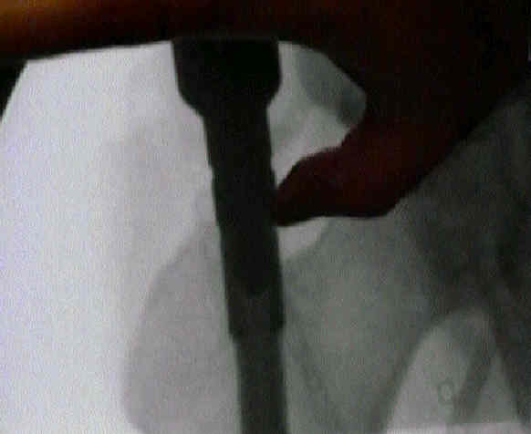
- with difficult passage of the guidewire across of the frx site it is accetpable to make a small lateral incision over the frx site, which allows passage
of the surgeon's gloved finger to palpate and reduce the fracture site.
- references:
- A vascular complication of trochanteric-entry femoral nailing on a fracture table
- Clamp-Assisted Reduction of High Subtrochanteric Fractures of the Femur: Surgical Technique
- Fracture Reduction:
- ref: Techniques of Obtaining and Maintaining Reduction During Nailing of Femur Fractures
- Determination of Leg / Nail Length: 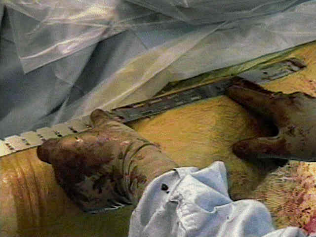
- appropriate nail length is determined by direct measurement of depth of the guide wire insertion;
- non-comminuted frx:
- w/ non comminuted frx it is assumed that leg length descrepancy is not an issue, and therefore it is important
to maximize frx opposition;
- once the nail is driven across the frx site, release traction and apply compress the frx fragments (by pushing
the distal fragment proximally), while the nail is driven distally;
- comminuted frx:
- w/ marked comminution, measure the uninjured side against nails of known length under the image intensifier, and then adjust
traction on the injured side to fit this predetermined nail length;
- once proper leg length has been determined, it is important to stick with the measured nail length, rather than compressing the fracture
ends which can lead to gross shortening at the frx site;
- the nail is driven into the distal fragment to the appropriate depth and then the distal interlocking screws are inserted;
- subsequently traction is applied while the nail continues to be driven in with the slap hammer until proximal end of the
nail reaches greater trochanter;
- ref: Assessing Leg Length After Fixation of Comminuted Femur Fractures
- Nail Insertion Down Medullary Canal:
- insert nail to frx site, & use it to assist w/ reduction;
- with trochanteric entry, there may a lateral to medial direction of the nail within the canal, which can lead to fracture comminution
as the nail is driven across the fracture site;
- consider rotating the nail 90 deg so that the apex is medial (points the nail tip lateral), which allows the nail to cross the fracature
site in a colinear direction without fracture site comminution;
- once the nail is across the fracture, the nail can then be re-rotated into an anatomic position;
- once nail is across the fracture, release traction and apply manual compress across the frx site as the nail is driven distally;
- achieve rotational alignment; (afterwards, don't rotate distal frag);
- as nail is driven down the distal frag, its important to shift the distal fragment either laterally or medially to avoid varus or valgus deformity at frx site;
- if proximal end of nail is below tip of greater troch, then use proximal end cap (can be extended up to 20 mm)
- Distal Interlocking;
- may be performed prior to proximal interlocking;
- it is important not to rotate the leg to obtain a perfect circle, rather, the flouro machine should be rotated to obtain a perfect circle;
- the former will result in a malrotation deformity of the leg;
- dynamization
- Fracture Rotational Alignment and Fracture Compression:
- release traction;
- rotational alignment: ensure that knee and foot are in proper rotational alignment;
- manually compess distal frag to the proximal fragment;
- references:
- Clinical determination of femoral anteversion. A comparison with established techniques.
- Rotational malalignment after intramedullary nailing of femoral fractures.
- The effect of rotational deformity on patellofemoral parameters following the treatment of femoral shaft fracture
- A forward-striking technique for reducing fracture gaps during intramedullary nailing: A technical note with clinical results.
- Proximal Inter-Locking;
- gently tap the trochar assembly to cortex surface, but do not penetrate cortex)
- reference:
- Cephalomedullary screws as the standard proximal locking screws for nailing femoral shaft fractures.
- Femoral anteversion
- Post Operative Care:
- patients are generally kept either touch down or partial wt bearing depending on the stability of the fracture;
- in the experimental report by Brumback RJ, et al (1999), the authors found that statically locked nails should provide enough stability (even
in unstable fractures) to allow protected wt bearing as tolerated;
- reference:
- Immediate Weight-Bearing After Treatment of Comminuted Fracture of Femoral Shaft with Statically Locked intrameduallary Nail.
- Complications of IM Nails: 
- compartment syndrome of thigh
- infected IM nails:
- fat embolism syndrome
- Mortality after reamed intramedullary nailing of bilateral femur fractures.
- avascular necrosis from IM nailing:
- broken nail
- Endomedullary Rods in Long Bones: A Danger Aboard
- non union: (see general discussion of non union)
- in the report by Weresh MJ, et al, the authors noted that a significant number of patients undergoing reamed exchange nailing of femoral
shaft non unions required additional procedures to achieve fracture healing;
- they noted that exchange nailing by itself may not be sufficient for fracture healing;
- ref: Failure of exchange reamed intramedullary nails for ununited femoral shaft fractures.
- in the report by Bellabarba C, et al, the authors report on a consectutive series of 23 femoral non unions
of femoral shaft fractures treated by previous IM nailing;
- surgical treatment consisted of indirect fracture reduction techniques using 95 deg condylar blade plate;
- 21 of 23 non unions healed without further intervention (two other fractures had hardware failure);
- refs:
- Results of indirect reduction and plating of femoral shaft nonumions after intramedullary nailing.
- Distal femoral nonunion treated with interlocking nailing.
- References:
Abduction Strength Following Intramedullary Nailing of the Femur.
The anatomy and functional axes of the femur.
Placing Femoral Intramedullary Nails in Severely Bowed Femurs