

- Discussion:
- transverse carpal ligament, is a heavy band of fibers which runs between hamate & pisiform medially to scaphoid and trapezium laterally, and forms
fibrous sheath which contains carpal tunnel anteriorly within fibro-osseous tunnel;
- posteriorly, tunnel is bordered by carpal bones, and transports median nerve & finger flexor tendons from forearm to hand;
- lies deep to palmaris longus & is defined by 4 bony prominences;
- proximally, by pisiform & tubercle of scaphoid;
- distally by hook of hamate & tubercle of trapezium;
- from hamate & pisiform medially to scaphoid & trapezium laterally;
- transverse carpal ligament, portion of volar carpal ligament, runs between these 4 prominences & forms fibrous sheath which contains
carpal tunnel anteriorly w/in fibro-osseous tunnel;
- posteriorly tunnel is bordered by carpal bones;
- Superficial Anatomy:
- palmaris longus passes in front of flexor retinaculum to become continuous with the palmar fascia (see transverse carpal ligament);
- palmar cutaneous branch of median nerve, which innervates skin over base of thenar eminence, arises short distance prox to flexor
retinaculum, pierces deep fascia, & course superficial to the flexor retinaculum to reach the skin;
- palmar cutaneous nerve of ulnar nerve course superficial to transverse carpal ligament & is not involved in CTS;
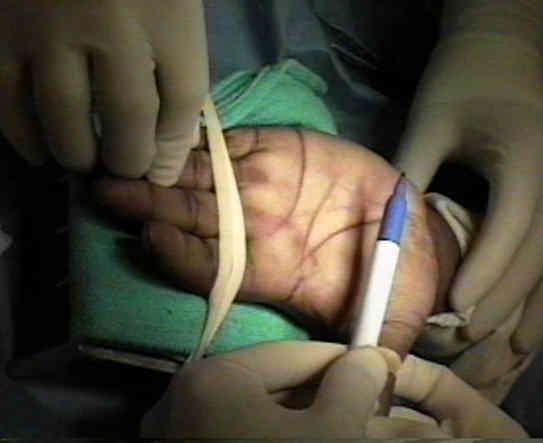
- distal volar flexion crease crosses proximal end of scaphoid and pisiform & marks proximal edge of TCL;
- references:
- Safety of carpal tunnel release with a short incision. A cadaver study
- Anatomy of neurovascular structures around the carpal tunnel during dynamic wrist motion for endoscopic carpal tunnel release
- Contents of Tunnel:
- tunnel transports median nerve & finger flexor tendons (FDS, FDP , & FPL);
- motor branch of median nerve in hand arises under or just distal to flexor retinaculum, & winds around distal border of retinaculum
to reach hypothenar muscles and the lateral 2 lumbricals;
- numerous variations in the branching have been described;
- sensory branches innervate lateral three and 1/2 digits & palm of the hand;
- references:
- Anatomic variations of the median nerve in carpal tunnel release
- Anatomical relationships among the median nerve thenar branch, superficial palmar arch, and transverse carpal ligament
- Guyon's Canal
- ulnar nerve & artery do not pass thru tunnel but lie superficial to it in guyon's canal
- pisiform is palpable & serves to mark entry, on its lateral aspect, of ulnar nerve and artery into the hand;
- Landmarks:
- distal volar flexion crease crosses proximal end of the scaphoid & pisiform & identifies proximal edge of the transverse carpal ligament;
- pisiform is palpable and just laterally will identify entry of ulnar nerve and artery into hand;
- all thenar & hypothenar muscles, except the abductor minimi, originate partly from the transverse carpal ligament.
- The Palmar Fat Pad Is a Reliable Intraoperative Landmark During Carpal Tunnel Release
- Kaplan's Line:
- Kaplan oblique line (line drawn from apex of interdigital fold between thumb and index finger, toward ulnar side
of hand, parallel w/ proximal palmar crease, & passing 4-5 mm distal to pisiform bone
The transverse carpal ligament. An important component of the digital flexor pulley system.
Transverse carpal ligament reconstruction in surgery for carpal tunnel syndrome: a new technique.
Anatomy of the flexor retinaculum.
Prevalence of anatomic variations encountered in elective carpal tunnel release