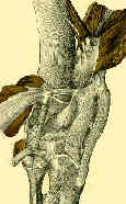
 - Anatomy:
 - origin:
- lateral condyle & posterior horn of lateral meniscus fibular head;
- popliteus muscle has 3 origins, strongest of which is from lateral femoral condyle, just anterior and inferior to the LCL origin;
- another origin is from fibula and from the posterior horn of the lateral meniscus;
- femoral and fibular origins form the arms of an oblique Y shaped ligament, the arcuate ligament;
- popliteus evolution:
- fibula originally articulated with the femur with the popliteus tendon inserted on the fibular head;
- w/ subsequent distal migration of the fibular head, required that the popliteus tendon achieve a
femoral attachment while keeping the original fibular head insertion;
- insertion:
- posterior surface of tibia above soleal or popliteal line;
- popliteus muscle arises from the proximal 10 to 12 cm of the tibia
- popliteus tendon runs deep to LCL and passes thru a hiatus in the coronary ligament to attach to the femur at a point anterior and distal
to the femoral attachment of LCL;
- nerve supply: tibia, L4, L5, S1; (see innervation)
- action:
- unique w/ a reversal of its origin and insertion w/ proximal attachment (origin) is its tendinous portion while its muscle belly sits distally;
- its primary f(x) is internal rotation of knee, unlocking of knee being accomplished by virtue of contourof articulation & retracting of posterior aspect of lateral meniscus;
- rotates the tibia medially on the femur & femur laterally on the tibia, depending on the one fixed;
- in a minority of pts, it withdraws the meniscus during flexion, and provides rotatory stability to the femur on the tibia;
- helps to brings the knee out of the position of full extension;
- helps with posterior stability of the knee in preventing posterior translation of the tibia on the femur;
- prevents excessive external rotation of the tibia during knee flexion from 20 - 130 deg,
- resists excessive varus rotation of the tibia during flexion from 0 - 90 deg
- Popliteus Tendonitis: (see running injuries)
- popliteus tendon courses from the proximal tibia to the distal femur, passing beneath the fibular collateral ligament;
- popliteus tendinitis is most easily detected with the leg in a figure of four (cross legged) position and then palpating just posterior and just anterior to the LCL ligament
The popliteus tendon
Popliteus tendon rupture. Case report and review of the literature.
Popliteus tendon tenosynovitis.
Avulsion of the popliteus tendon: a rare cause of chondral fracture and hemarthrosis.
The popliteus tendon and its fascicles at the popliteal hiatus: gross anatomy and functional arthroscopic evaluation with and without anterior cruciate ligament deficiency.
Arthroscopic Evaluation of the Popliteus: Clues to Posterolateral Laxity
Location and classification of popliteus tendon’s origin: cadaveric study
Popliteus Tendon Resection During Total Knee Arthroplasty: An Observational Report
Femoral Radiographic Landmarks for Popliteus Tendon Reconstruction and Repair. A New Method of Reference

