

- See: dorsal barton's frx and dorsal approach to the wrist:
- Surgical Approach:
- patient is supine w/ wrist slightly flexed & placed on arm board;
- perform a provisional reduction before the incision is made;
- Z or S shaped incision from base of second metacarpal over wrist to distal forearm;
- alternatively, make a straight midline longitudinal one in line w/ third metacarpal and into the distal forearm;
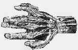
- the incision will be made through the 3rd compartment;
- this passes safely between the dorsal sensory branches of the ulnar nerve medially, and ofthe radial nerve laterally;
- extensor retinaculum between 3rd and 4th extensor compartments is reflected off of wrist capsule w/ care to avoid any damage to capsule;
- alternatively, incise directly over the EPL tendon sheath, which will completely avoid injury to the 4th compartment;
- some surgeons, will step cut the distal portion of the extensor retinaculum (from the first compartment to the fourth compartment) so that the retinaculum can rest between the plate and the overlying tendons;
- EPL is mobilized out of its sheath and is reflected radially;
- subperiosteally elevate the fourth compartment, w/o violating the tendon sheath or wrist capsule;
- place Homan retractors on either side of the radius;
- longitudinally incise thru the dorsal capsule in line w/ Lister's tubercle, and then elevate the wrist capsule off of the dorsal rim of the distal radius including the dorsal radiotriquetral ligament;
- wound closure:
- some surgeons will, remove Lister's tubercle and will leave the EPL out of its tunnel;
- retinaculum is approximated with the EPL left dorsal to the retinaculum;
- Limited ORIF: Displaced Lunate Fossa:
- reduction may be facilitated by ligamentotaxis using external fixation;
- radial styloid process and scaphoid facet are more amenable to reduction thru ligamentotaxis or by manipulation and reduction w/ large, pointed bone clamp;
- incongruity often remains in the lunate facet of the distal radius;
- consider combining external skeletal fixation w/ open reduction of displaced lunate facet through small, longitudinal dorsal incision and elevation of impacted fragment w/o direct visualization of the surface of joint
- support reduction with transverse or oblique K wires & bone graft;
- watch for settling of elevated articular fragments;
- external fixation frame can be removed by six weeks after application;
- references:
- Limited open reduction of the lunate facet in comminuted intra-articular fractures of the distal radius.
- Treatment of severely comminuted intra-articular fractures of the distal end of the radius by open reduction and combined internal and external fixation.
- Plate Fixation: (see plate fixation techniques)
- attempt to interpose a soft tissue layer between the plate and extensor tendons;
- consider a step cut division of the extensor retinaculum, so that the distal half of the retinaculum can be interposed between the plate and tendons;
- EPL is prone to rupture from rubbing over plate & screws;
- if there is > 4-5 mm of impaction, bone graft from iliac crest or an allograft is recommmedned to fill metaphyseal defect;
- application of plate requires removal of Lister's tubercle, and subperiostral exposure of the fracture;
- distal comminution precludes use of screws in distal fragment;
- complications:
- in the study by Ring, et al (1997), 5/22 patients who underwent ORIF w/ a low profile plate developed extensor tendinitis;
- authors of this study suggest the inclusion of a retinacular flap to be placed between the 2nd compartment extensor tendons and the overlying plate;
- Prospective multicenter trial of a plate for dorsal fixation of distal radius fractures.
- Post Operative Care:
- doral plate should be removed following fracture healing (to minimize tendon irritation);
Treatment of displaced intra-articular fractures of the distal end of the radius with plates.
Prospective multicenter trial of a plate for dorsal fixation of distal radius fractures.