

- See: Bone Tumor Menu
- Discussion:
- an osteochondroma that results from a dysplasia of peripheral growth plate;
- as its name implies, multiple hereditary exostosis is an inherited condition which produces multiple exostoses;
- occurs in 1 out of 50,000 people;
- typically inherited as an autosomal dominant trait, w/ 96% penentrance;
- 10% may have no family history;
- EXT1 on 8q24.1 and EXT2 on 11p13 are the two genes most strongly associated with MHE
- ref: EXT1 regulates chondrocyte proliferation and differentiation during endochondral bone development.
- average age of diagnosis is 3 yrs;
- cartilaginous exostoses arise from metaphyses, point away from epiphysis, and appear to extend down diaphysis during growth;
- they increase in size & number w/ growth, but may become latent at maturity;
- osteochondromas are seen in several sites;
- in over 90% of cases distal tibia, proximal tibia, proximal femur, and proximal humerus are involved.
- will also involve iliac crests, scapulae, and ribs;
- sarcomatous degeneration:
- chondrosarcoma may develop in 1-2% of patients > 21 yrs of age;
- lesions at risk are those occurring near pelvis, scapula, proximal humerus, proximal femur, & spine have increased risk of malignant transformation;
- change in size of the exostosis or onset of pain in an affected adult is cause for concern and investigation.
- monitoring pts via annual bone scans has been recommended, but its efficacy remains unproven.
- Radiographic Distribution of Lesions:
- scapula: 40%
- proximal humerus: 50%
- ribs: 40%
- distal radius: 30%
- distal ulna: 30%
- hands: 20-30%
- ilium: 15%
- mid femur 30%
- distal femur: 70%
- proximal tibia 70%
- proximal fibula 30%
- distal tibal and fibula: 20-25%
- feet: 10-15%
- Clinical Presentation:
- despite the high frequency of radiographic findings, a much smaller percentage of patients will demonstrate clinical lesions;
- 75% of patients will have at least one clinically evident lesion;
- 50% will demonstrate a forearm deformity;
- 45% will demonstrate an ankle deformity;
- 20% will demonstrate knee deformity;
- clinical problems include:
- fracture;
- pressure of exostosis on surrounding soft tissues;
- neurovascular compromise;
- stature:
- 40% will demonstrate short stature;
- individuals may be small, but are not considered dwarfs;
- leg length inequality may be severe enough to require equalization procedures in half of the individuals;
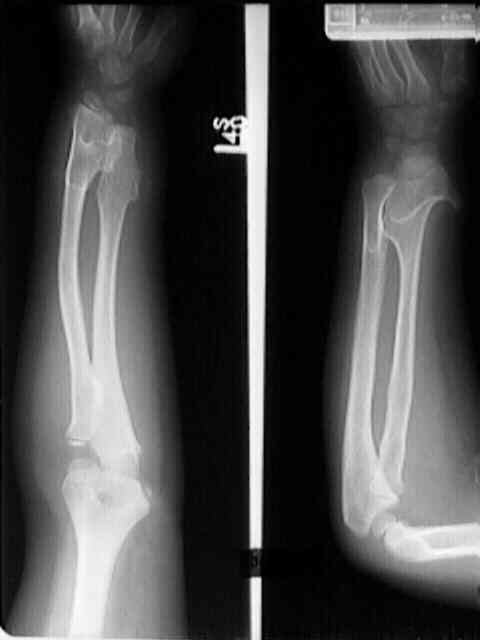
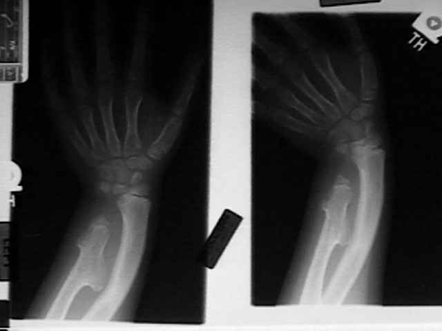
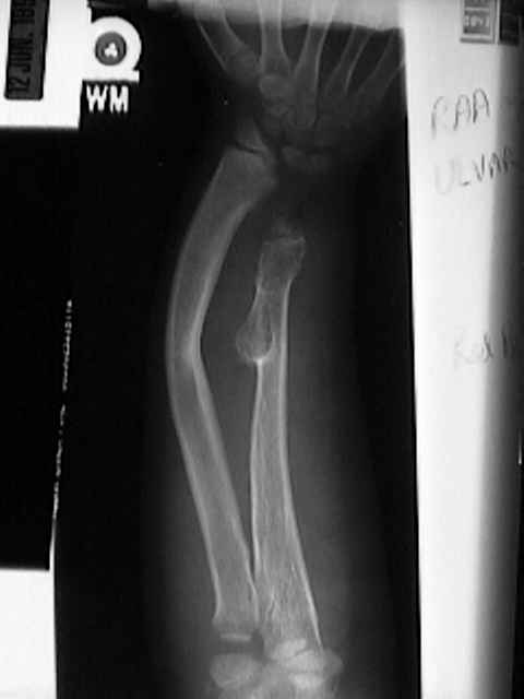
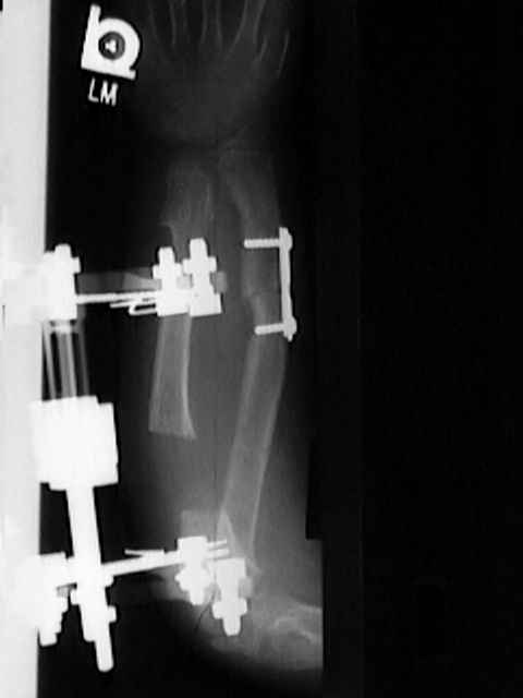
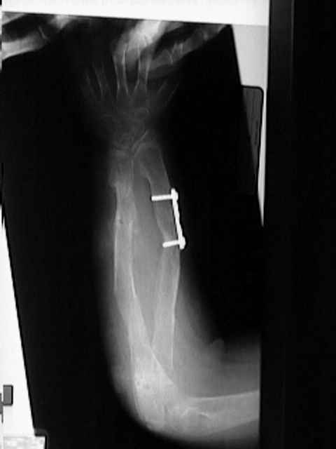
- Ulna in MED:
- shortening is the major problem.
- distal ulna contributes more to total bone length than does distal radius and therefore is more prone to shortening from a distal exostosis;
- over 50% of patients will be affected;
- diff dx: multiple enchondromatosis:
- effects of ulnar shortening include;
- radial bowing and angulatory deformity;
- ulnar deviation of the wrist;
- ulnar translation;
- radial head dislocations
- more common w/ severe ulnar shortening;
- may lead to loss of pronation and increased ulnar variance;
- ulnar varience is measured as the difference between the radial and ulnar physis;
- treatment:
- probably most patients with forearm involvement do not require treatment despite severe deformity;
- references:
- Evaluation of the Forearm in Untreated Adult Subjects with Multiple Hereditary Osteochondromatosis
- Forearm Deformities in Hereditary Multiple Exostosis: Clinical and Functional Results at Maturity
- excision of a distal osteochondroma gives unpredictable results;
- radial head should never by excised in a growing patient;
- stapling of radial hemiepiphysis may improve wrist alignment as well as improving supination and pronation;
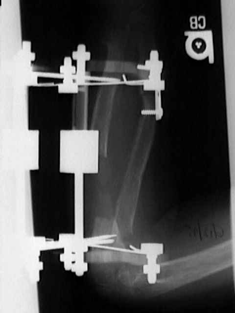
- ulnar lengthening:
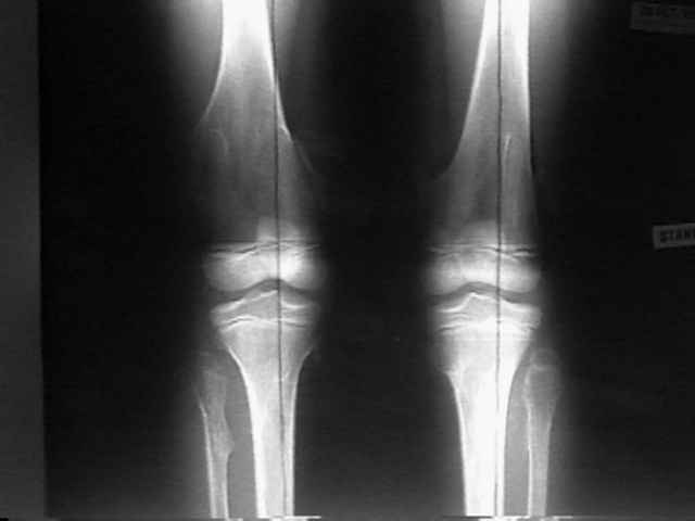
- Knee Joint in MED:
- leg length inequality may require equalization procedures in 50%;
- fibular involvement by osteochondroma may cause: genu valgum;
- Ankle in MED:
- valgus ankle deformity;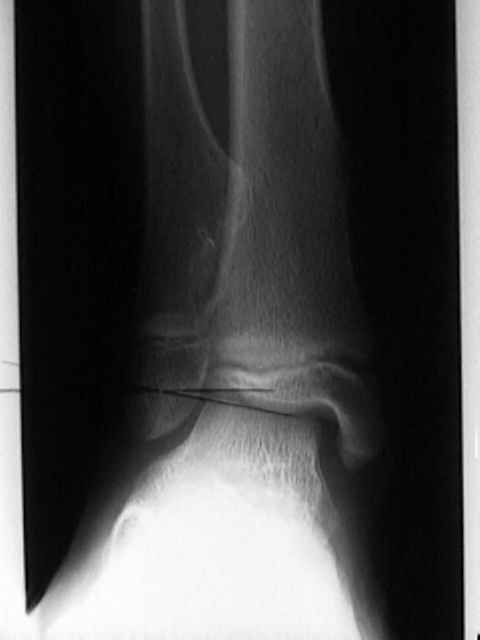
- diastasis of the ankle;
- surgical indications:
- pain from trauma or from prominent masses;
- ankle pain assoc w/ deformity
- w/ tibiotalar valgus > 15 deg, hemiepiphyseal stapling is indicated;
- surgical treatment:
- fibular lengthening + hemiepiphyseal stapling when valgus coexists w/ leg length discrepancy
Multiple hereditary osteochondromata.
Management of deformities of the forearm in multiple hereditary osteochondromas.
Synovial osteochondromatosis: a histopathological study of thirty cases.
Deformities of the forearm in patients who have multiple cartilaginous exostosis.
The natural history of hereditary multiple exostoses.
Knee deformities in multiple hereditary exostoses. A longitudinal radiographic study.
Hereditary Multiple Exostoses: One Center’s Experience and Review of Etiology.
Involvement of the Spine in Patients with Multiple Hereditary Exostoses