

Discussion
- although a traumatic etiology is believed to play a major role in production of these lesions, idiopathic osteonecrosis may be another factor;
- anterolateral lesions:
- may result from impaction of talus on fibula as the dorsiflexed ankle is forced into inversion (see ankle sprain);
- the vast majority are caused by trauma;
- these lesions tend to be shallow;
- posteromedial lesions:
- most of these lesions probably arised from trauma (but many will have an atraumatic etiology);
- may result from impaction of posteromedial talus on tibia, as plantar flexion ankle is forced into inversion and exteranal rotation;
- these lesions are deeper and cup shaped;
- exam: palpate just posterior to the medial malleolus with the ankle dorsiflexed (may resemble pending rupture of posterior tib)
Exam
- joint line tenderness and effusion;
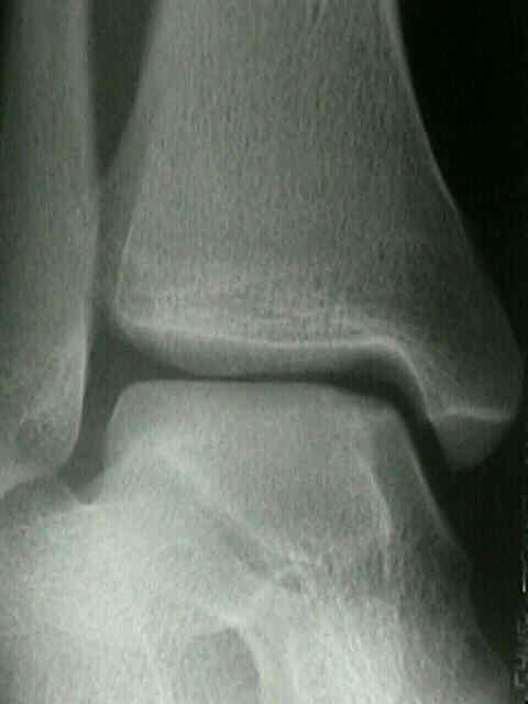
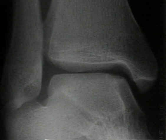
Radiographs
- osteochondral frx may be anterior or posterior to dome, requiring plantar or dorsiflexion of ankle to be visible on mortise view;
- if radiographs are negative consider repeat radiographs in 2-4 weeks;
- radiographic classification: (Berndt and Harty)
- note: that radiographic findings may or may not correlate w/ arthroscopic findings nor prognosis;
- I: small area of compression;
- II: partially detached osteochondral lesion;
- III: completely detached, non-displaced fragment;
- IV: detached and displaced fragment;
- note: that radiographic findings may or may not correlate w/ arthroscopic findings nor prognosis;
Bone Scan
- usually not ordered until 8-12 weeks following diagnosis;
- a negative bone scan will r/o the diagnosis;
- if bone scan is positive then order either CT or MRI;
CT Scan
- offers more accurate staging of the lesion;
Treatments
Non Operative Treatment
- no evidence that non wt bearing cast offers improved results over wt bearing casts;
- no evidence that patients need to be immobilized if they are kept non wt bearing;
Operative Treatment
For a comprehensive review of Osteochondral grafting on Wheeless readers should also visit:
Osteochondral Lesions of the Talus – Allograft Repair
- Arthroscopy of the Ankle:
- osteochondral lesions of the talus can be debrided, and loose bodies and small osteochondral fragments can be removed;
- use of non-invasive or invasive distraction improves access to joint and allows adequate debridement and curettage of bed;
- anterolateral lesions can be addressed from the anterolateral portal;
- in the study by Kumai, et al (1999), the authors noted good clinical results w/ arthroscopy and K wire drilling of the OCD lesions in patients who were younger than 50 years;
- posteromedial lesions can be difficult to access;
- w/ large fragments ORIF may be required, w/ osteotomy of the medial malleolus being required for exposure;
- ORIF allows direct observation of the lesion and accurate repair;
- w/ ORIF immobilization in a non wt bearing cast is required for 6-10 weeks
References
- Osteochondral fractures of the talus. A long-term follow-up.
- Osteochondral fractures of the dome of the talus.
- Osteochondral lesions of the talus.
- Osteochondritis dissecans of the dome of the talus. Computed tomography scanning in diagnosis and follow-up
- Osteochondritis dissecans of the talus (transchondral fractures of the talus): review of the literature and new surgical approach for medial dome lesions.
- Arthroscopic treatment of osteochondral lesions of the talus.
- Surgical treatment of transchondral talar-dome fractures (osteochondritis dissecans). Long-term follow-up.
- Transchondral fractures (osateochondritis dissecans) of the talus.
- Arthroscopic management of transchondral talar dome fractures (osteochondritis dissecans) and anterior impingement lesions of the ankle joint.
- Osteochondral lesions of the talus.
- Arthroscopic Drilling for the Treatment of Osteochondral Lesions of the Talus
- Nonoperatively managed stage 5 osteochondral talar lesions.