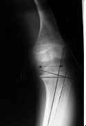
- See: Rickets:
- Discussion:
- this is the classic form of the disease;
- it usually results from a deficiency in dietary intake of vitamin D, often coupled with inadequate exposure to sunlight;
- reduced vitamin-D intake causes a diminution in the absorption of calcium from the GI tract;
- insufficient absorption results in a diminished serum calcium level, which subsequently causes a secondary hyperparathyroidism and resultant
increase of the serum Ca concentration to low normal and a phosphate diuresis;
- combination of a reduced amount of mineral (both ionized Ca & phos) & secondary hyperparathyroidism is presumed to be principal factor
responsible for pathological changes in epiphyseal plates (in rickets) & bones & resultant alterations in x-rays images;
- chelators in the diet:
- see: GI and biliary causes of rickets
- major dietary inclusions that can bind calcium and render it unabsorbable in the ionized state are phytate (from some coarse cereals), oxalate (present
principally in spinach), & excess of phosphate;
- phosphorus deficiency:
- can occur with the introduction into diet of beryllium (as an industrial toxic material) or, much more commonly, aluminum (present as Al(OH) in
many antacid preparations);
- infantile nutritional rickets:
- most common in african americans who are breast fed for more than 6 months and who do not receive vitamin D;
- Clinical Findings:
- children with this disorder usually show the abnormality by age of one year or, in florid cases, even earlier.
- they may display severe weakness, inability to walk, and remarkable deformities of the skeleton (genu varum and swollen wrists);
- adults, in contrast, have few localizing symptoms or findings;
- they may complain of malaise, easy fatigability, bone pain & tenderness, weakness, or a combination of these symptoms;
- physical findings in the adult are sparse but may include a Trendelenburg gait and tenderness over osseous prominences;
- great concern, particularly in dealing w/ elderly pt, is that osteopenia or fracture may be attributed to osteoporosis when, in fact, it represents
nutritional osteomalacia;

- Radiographs:
- Labs:
- hypophosphatemia may be present due to phosphate diuresis (may be normal in some patients);
- hypocalcemia may be present (or may be low normal);
- alkaline phosphatase is generally elevated;
- BUN and Cr are normal

