

- Discussion:
- used to assess impingement syndromes (coronal oblique views) and less often glenoid pathology (transaxial views);
- it accurately identifes full-thickness rotator cuff tears;
- defects show up with high signal intensity traversing supraspinatus tendon on T2 images;
- MRI is less specific in diagnosing tendinitis & partial tears;
- external rotation:
- patients should keep the shoulder in external rotation during the exam;
- position keeps slight tension on anterior capsular structures;
- external rotation permits maximum visualization of the supraspinatus insertion, and prevents confusing overlap with the
infraspinatus tendon on coronal oblique images;
- signal intensity characteristics:
- fat suppressed T2 weighted:
- long repitition time and long echo time
- water gives bright signal and fat gives very dark signal;
- allows evaluation of marrow pathology;
- STIR images:
- long repitition time and variable echo time;
- water gives bright signal and fat gives very dark signal;
- allows evaluation of marrow pathology;
- gradient echo:
- short repitition time and short echo time
- intermediate fat signal and intermediate to bright water signal;
- for evaluation of articular cartilage, blood, and PVNS;
- proton density:
- long repitition time and short echo time;
- intermediate to high fat signal and intermediate water signal;
- high resolution for evaluation of labral tears, but poor evaluation of marrow;
- references:
- Rotator Cuff: Evaluation with US and MR Imaging.
- Labral injuries: accuracy of detection with unenhanced MR imaging of the shoulder.
- Specific Views: (see Anatomy of the shoulder (MR) - Atlas of the human body)
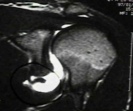
- transaxial view of the shoulder:
- evaluates shoulder capsule, glenoid labrum, subscapularis, biceps and for evaluation of a Hill Sachs Lesion;
- protocols: fat suppressed T2 wt images and proton density weighted images
- oblique saggital 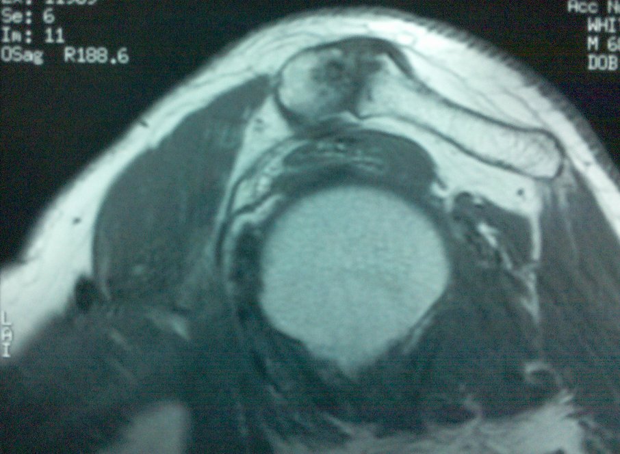
- plane is perpendicular to supraspinatus;
- non fat suppressed T1 wt images and fat suppressed T2 wt images;
- acromial morphology is best evaluated on sagittal oblique magnetic resonance images;
- (see radiographic evaluation of impingement syndrome)
- useful for imaging the subscapularis;
- w/ subscapularis tears look for disruption of the transverse ligament;
- biceps tendon is followed from medial to lateral as it courses from its intra-articular origin on supraglenoid
tubercle to its extracapsular location in the bicipital groove laterally;
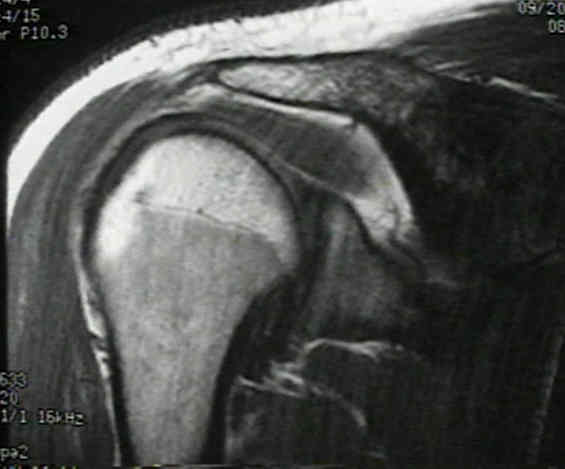
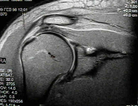
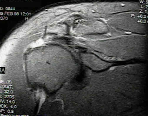
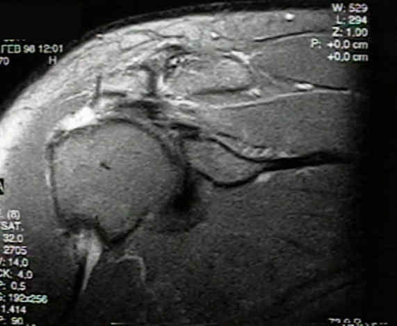
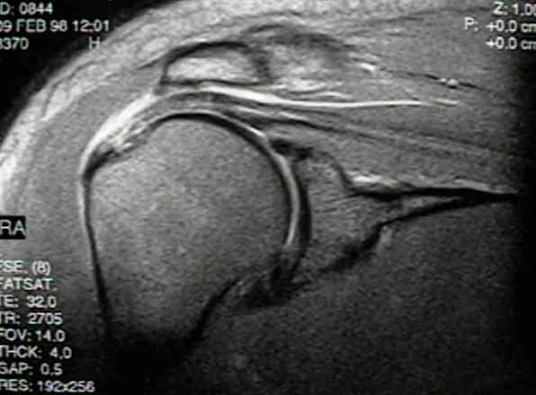
- coronal oblique views of the MRI:
- protocols: fat suppressed T2 wt images and proton density weighted images
- Hagl Lesion:
- avulsion of inferior ligament from the humerus;
- references:
Humeral avulsion of the anterior shoulder stabilizing structures after anterior shoulder dislocation: demonstration by MRI and MR arthrography.
Magnetic resonance imaging of the shoulder. Sensitivity, specificity, and predictive value.
Magnetic resonance imaging of the shoulder.
Shoulder instability: evaluation with MR imaging.
Abnormal findings on magnetic resonance images of asymptomatic shoulders.
The use of MRI about the shoulder. Beltran J. J Shoulder Elbow Surg. 1992;1:321.