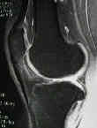
- Discussion:
- most important portal, initial site for scope insertion;
- structures that are difficult to see:
- PCL
- anterior portion of lateral meniscus
- posterior horn of medial meniscus may not be adequately seen;

- Portal Placement:
- knee should have already been injected w/ lidocaine/marcaine inorder to distend the joint;
- the needle can be used to "sound" the joint;
- knee is flexed 30 deg in order to engage patella into trochlear groove and to keep tension on the retinaculum;
- portal is placed at level of inferior and lateral edges of patella;
- portal is located at least 1 cm above the lateral joint line and approximately 1 cm lateral to the margin of the patellar tendon;
- some surgeons always place this portal just below the most inferior level of the patella;
- the superior portion of the meniscus is vulnerable during suprameniscal portal placement;
- by keeping the portal slightly off the edge of the patellar tendon, the fat pad will be avoided, however, this will make visualization of the posterior horn of the medial meniscus and lateral intercondylar notch even more difficult;
- when using knife, the blade should be directed horizontally with the blade directed away from the patella tendon;
- once the blade is buried beneath the skin, the blade can be rotated upwards 90 deg in order to gently incise the capsule vertically (hence the scope will not be fighting the capsule during the case);
- bring the blade back to its horizontal position and as it is removed, gently extend the incision by another 2 mm (if the skin and capsule are too tight the surgeon will struggle, and if the incisions are too large excessive fluids will be lost and the capsule will distend poorly;
- arthroscope sheath w/ its blunt trocar is inserted thru portal & aimed toward intercondylar notch;
- ACL reconstruction:
- w/ ACL recon, need to place portals close to patellar tendon to allow instrumentation of notch;
- keep this portal slightly higher to avoid fat pad;
- when intentionally placing the portal slightly higher than usual, mark out the level of the posterior joint line to ensure that access for posterior mesical repair is possible;
- usually this means that the scope should not be angled more than 15 deg off the slope of the tibial joint surface;
- Portal Too Lateral:
- makes visualization of the intercondylar notch difficult;
- may cause deflection of the scope by the lateral femoral condyle makes it difficult to move the leg to the figure of 4 position;
- Portal Too Low:
- if portal is placed too near joint line, anterior horn of lateral meniscus may be lacerated or otherwise damaged.
- also a low portal is more likely to enter fat pad;
- if the portal is being intentionally placed slightly lower than usual (as might be done for meniscal repair) keep the portal slightly off the margin of the patellar tendon (to avoid the fat pad);
- Portal Too High:
- high anterolateral portal:
- if placed too superiorly to joint line, does not permit scope to enter the space between the femoral and tibial condyles
- prevents visualization of the posterior horns of menisci;
- however, a more superior portal placement avoids fat pad;
- Portal Too Medial:
- placement of arthroscope immediately adjacent to edge of patellar tendon results in possible penetration of the fat pad, causing difficulty in viewing & maneuvering arthroscope w/in joint;
- fat pad penetration is more likely w/ low portal placement;
- Misc:
- fat pad:
- when fat pad appears prominent and is obstructing visualization, resist the urge to shave away the fat pad;
- instead, attempt to "push and pull" the scope either into the notch or the supra-condylar pouch inorder to pull the fat pad anteriorly out of the field of vision

