(see also: Elbow Arthritis)
Equipement
- 30 deg, 4 mm arthroscope;
- arthroscopic pump;
Positioning
- patient is usually prone with sandbag placed under antecubital fossa;
- TV monitor is positioned opposite of the patient;
- ulnar nerve is palpated to pinpoint its location and to ensure that it does not subluxate with elbow flexion
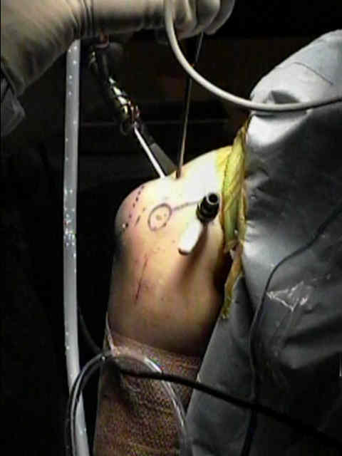
- instill fluid into the joint thru the aconeus triangle;
- through the lateral soft spot, which is bound by the radial head, lateral epicondyle, and olecranon
- when the elbow is distended, the major neurovascular structures are positioned farther away from the portal sites
Portal Placement
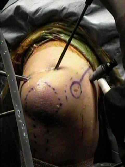
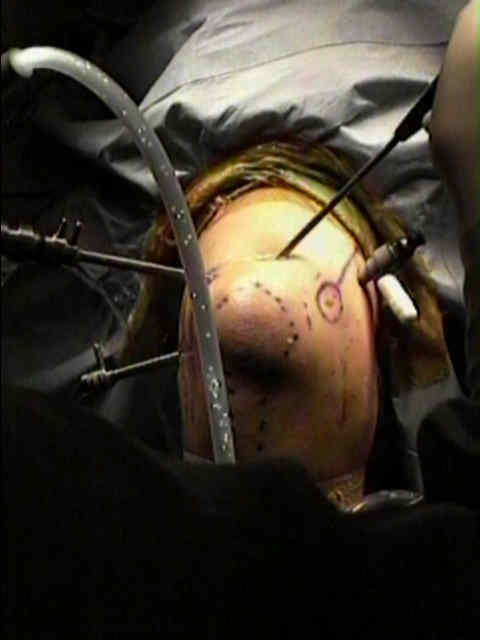
posterolateral portal
- portal is located thru the center of the aconeus triangle;
- when the anterior aspect of the joint is being visualized, the posterolateral portal can be used as an outflow portal;
- posterolateral portal can be used to visualize the posterior elbow structures including the olecranon fossa;
- use of a 70 deg arthroscope facilitates visualization of the radiocapitellar joint;
- this portal allows debridement of the capitellum (in the case of osteochondritis dissecans);
anterosuperior lateral
- considered safer than anterolateral portal;
- portal allows visualization of the ulnohumeral articulation, anteromedial aspect of elbow, coronoid fossa and process, and anterior
aspect of radiocapitellar joint; - portal is placed 2 cm superior to the anterior aspect of lateral epicondyle;
- blunt dissection into the joint capsule helps to avoid injury.
cautions
- PIN is most at risk, (less than the anterolateral portal) and lateral antebrachial cutaneous nerves are at risk;
anterolateral portal
- this portal can be used for instrumentation as well as visualization of the lateral aspect of the radial head;
- this portal is often established first;
- w/ elbow flexed 90 deg, the portal is located 3 cm distal and 1-2 cm anterior to the lateral epicondyle, which should bring portal just anterior and proximal to the radial-capitellar articulation w/ the portal driven toward the center of the trochlea;
- elbow is kept flexed during trochar insertion since extension brings the radial nerve closer to the joint (3 to 7 mm);
hazards
- the portal pass through the ECRB and supinator;
- posterior interosseous nerve is at risk w/ this portal, as it runs about 0.5 cm to 1 cm anterior and medial to the portal;
- in order to protect the PIN, this portal is made from inside outward under visualization from the proximal-medial portal;
- the scope is advanced anterolaterally across the radial head w/ care not to deviate too anteriorly;
- the light of the arthroscope will be visible thru the skin which will facilitate proper skin incision;
proximal anterolateral portal
- located 2 cm proximal and 1 cm anterior to the lateral epicondyle;
- this portal is significantly farther (on average 13.7 mm) from the radial nerve than other anterolateral portal sites;
- this portal allows for an excellent view of the anterior radiohumeral and ulnohumeral joints as well as the anterior capsular margin.
proximal antero-medial portal
- allows visualization of the following:
- anterior elbow including the anterior joint capsule, medial condyle, coronoid process, trochlea, capitellum, and the radial head;
- radial head is best visualized from the proximal anteromedial portal
- joint should already be distened w/ fluid;
- location is 2 cm proximal to medial epicondyle, and immediately anterior to the inter-muscular septum, using a longitudinal skin stab incision;
- ensure that the position of the intermusuclar septum is clearly demarcated;
- make 1/2 cm incision and spread w/ hemostat;
- trochar is inserted over the anterior surface of the humerus aiming for the radial head;
- maintain contact w/ anterior humerus at all times to reduce risk to N/V structures is minimized.
- trocar w/ it (arthroscopic sheath) is then inserted, followed by the scope;
- hazards:
- nerves at risk w/ this portal include the ulnar nerve, medial brachial cutaneous, medial antebrachial cutaneous, median nerve, and brachial artery;
- ulnar nerve lies 4 mm from this portal site;
- median nerve lies 7-20 mm away from portal with the elbow in flexion;
anteromedial portal
- some surgeons prefer to establish this portal first;
- elbow should be flexed 90 deg as the portal is established;
- placed 2 cm anterior and 2 cm distal to the medial epicondyle, placed under direct vision;
- the median nerve lies 1 to 2 cm anterior and lateral to this portal;
Complications: nerve damage
- be particularly careful in elbow that have altered anatomy or scarring (as from intra-articular fracture);
- this situation may distort normal landmarks, decrease arthroscopic distension, and cause nerves to remain adherent to capsule;
- in the report by Kelly EW, et al, the authors retrospective review of 473 consecutive elbow arthroscopies performed in 449 patients over an 18 year period was conducted;
- of the 473 cases, 414 were followed for more than six weeks;
- most common final diagnoses were osteoarthritis (150 cases), loose bodies (112), and RA or inflammatory arthritis (seventy-five);
- arthroscopic procedures included synovectomy (184), débridement of joint surfaces or adhesions (180), excision of osteophytes (164), diagnostic arthroscopy (154), loose-body removal (144), and capsular procedures such as capsular release, capsulotomy, and capsulectomy (73).
- a serious complication (a joint space infection) occurred after four (0.8%) of the arthroscopic procedures;
- minor complications occurred after fifty (11%) of the arthroscopic procedures;
- these complications included prolonged drainage from or superficial infection at a portal site after 33 procedures, persistent minor contracture of 20° or less after seven, and twelve transient nerve palsies (five ulnar palsies,
four superficial radial palsies, one posterior interosseous palsy, one medial antebrachial cutaneous palsy, and one
anterior interosseous palsy) in ten patients; - most significant risk factors for the development of a temporary nerve palsy were an underlying diagnosis of rheumatoid arthritis (p < 0.001) and a contracture (p < 0.05). There were no permanent neurovascular injuries, hematomas, or
compartment syndromes in our series, and all of the minor complications, except for the minor contractures, resolved
without sequelae.
- these complications included prolonged drainage from or superficial infection at a portal site after 33 procedures, persistent minor contracture of 20° or less after seven, and twelve transient nerve palsies (five ulnar palsies,
- references:
- Complications of Elbow Arthroscopy.
- Complete Transection of the Median and Radial Nerves During Arthroscopic Release of Post-traumatic Elbow Contracture.
- The course of the median and radial nerve across the elbow: an anatomic study
- Neurovascular anatomy and elbow arthroscopy: inherent risks.
- Elbow Positioning and Joint Insufflation Substantially Influence Median and Radial Nerve Locations
- Avoiding nerve damage during elbow arthroscopy.
- Prevention of Nerve Injury During Arthroscopic Capsulectomy of the Elbow Utilizing a Safety-Driven Strategy
- Arthroscopic surgery of the elbow. Therapeutic benefits and hazards.