

- See:
- Anterior Dislocation
- Posterior Dislocations:
- Discussion:
- note: w/ true AP of shoulder there should be no overlap of the humerus over the glenoid;
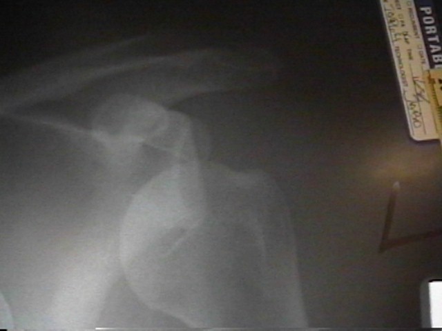
- Greater Tuberosity and Acromio Humeral Interval;
- AP in ext. rot. brings greater tuberosity into clear prominence;
- this x-ray taken w/ pts arm at side, hand supinated, & elbow extended;
- gives good visualization of the greater tuberosity;
- measurement of the acromiohumeral interval also can be done more accurately on the external rotation view;
- Calcific Tendonitis:
- AP w/ arm in internal rotation is taken with the arm at the side, elbow extended, and the forearm pronated;
- may also be helpful in calcific tendinitis, esp in the supraspinatus;
- AC Joint View: (see AC joint)
- AP w/ arm in 100 deg of abduction provides best view of AC joint;
- this projection allows the AC joint to clear the overlying scapular or acromial neck, so degeneration or osteophytes can be seen better;
- this view is a useful one in pts with true adhesive capsulitis;
- Technique:
- taken w/ pt's back flat on cassette & x-ray beam at right angles to this plane and centered on the shoulder;
- the scapula sits obliquely on the chest wall, and the glenoid surface is tilted approx. 35 to 40 deg anteriorly;
- the posterior aspect of the affect shoulder is placed up against the x-ray plate & opposite shoulder is rotated out approx. 40 deg;
- angled AP radiograph (body turned 35-40 toward cassette), shows glenoid fossa in profile
- Note Neck Shaft angle:
- angle created at intersection of lines that are perpendicular to anatomic neck and parallel to shaft of humerus;
- avg angle 143 deg (134 to 166) - angle is less in external rotation and angle is more in internal rotation