- See:

- Anterior Drawer Test
- Anatomy:
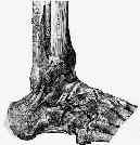
- it connects anterior fibula to neck of talus;
- ligament is thickening of ankle capsule that extends short distance from anterior edge of distal part of fibula to talar neck, anterior to its lateral articular facet;
- ligament is 20 mm long, 10 mm wide, and 2 mm thick;
- w/ the foot plantigrade, its fibers are oriented 75 deg to the floor;
- w/ plantar flexion, its fibers approach vertical orientation;
- anterior and posterior talofibular ligaments blend in with the joint capsule (unlike the calcaneal fibular ligament);
- thus capsular tears accompany tears of the 2 talofibular ligaments;
- distance from tip of fibula to center of fibular attachment of anterior talofibular ligament is 10 millimeters;
- Discussion:
- weakest of the lateral ligaments;
- prevents anterior subluxation of talus when ankle is in plantar flexion;
- orientation of ant talofibular lig depends on position of ankle joint.
- in plantar flexion, it is parallel to long axis of foot, whereas in dorsiflexion, it is aligned w/ tibial & fibular shafts;
- strain in ATFL is minimum in dorsiflexion & neutral, & it increased as ankle is moved progressively through plantar flexion;
- Exam:
- it is difficult to be certain which lig is being evaluated w/ each of clinical tests;
- commonly used tests for eval of lateral ligament laxity of ankle are anterior drawer & talar tilt tests.
- inversion (supination) test
- w/ ankle in plantarflexion: evaluates anterior talofibular ligament;
- in neutral / sl. dorisflexion: evaluates calcaneofibular ligament
The contribution of the anterior talofibular ligament to ankle laxity.
Biomechanical evaluation of the anterior drawer test: the contribution of the lateral ankle ligaments.
Instability of the hindfoot after lesion of the lateral ankle ligaments: investigations of the anterior drawer and adduction maneuvers in autopsy specimens.

