

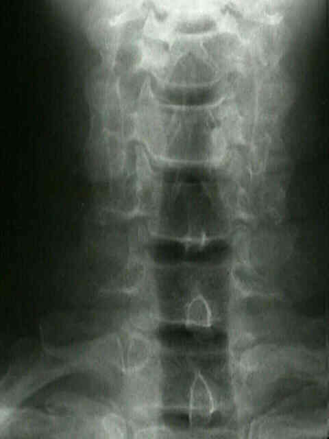
- Pillar View
- Discussion:
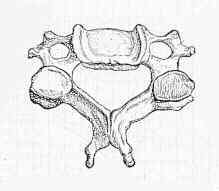
- this view demonstrates C3 thru C7 vertebral bodies, spinous processes and lateral masses;
- Evaluates:
- lateral mass fractures
- sagittal plane frxs (also called vertical compression frx) may be visualized on the anteroposterior view;
- this view may show altered separation between spinous process tips caused by flexion-induced injuries;
- signs of direct injury:
- malalignment of the spinous processes on the anterior view;
- lateral tilting of the vertebral body on the anterior view;
- because dislocation/sublux may be subtle on plain series any rotation of spinous processes on AP view should alert M.D. to exam oblique views where fascet relationships are best seen;
- saggital plane frx is verticle compression frx, but more specifically it is sagittally and not coronally oriented;
- this frx often occurs in combo with other fractures in the same or adjacent vertebrae, for example, laminar fracture, facet
dislocation, or teardrop fracture dislocation, extensive ligamentous damage, and paralysis;
- key feature is a midsagittal fracture plane extending from one vertebral end plate to other, which is best seen on AP view;
- lateral radiograph may show no abnormality at all;
- Radiographic Anatomy:
- 1st & 2nd vertebrae are obscurred in this projection by mandible and basiocciput, whereas lower cervical vertebrae & cervico-throracic
junction are well seen;
- lateral masses appear as bilateral smooth undualing margins, & spinous processes are in the midline;
- interspinous distances should be symmetric throughout;
- interspinous distance 1.5 times distance above or below level may indicate a dislocation or subluxation;
- unilateral facet dislocation may result in lateral rotation of one spinous process with respect to the others;
- Radiographic Technique:
- patient is erect or supine
- central beam is directed toward the C4 vertebra (at the point of Adam's apple) w/ 15-20 deg cephalic tilt;
- mandible is held open (open mouth anteroposterior) to see C-1 & C-2;
- in comatose pt, place gauze roll between teeth;
- shooting one view w/ beam slightly angulated cephalad and another w/ it slightly caudad increased likelihood of visualizing C-1
& C-2 well, especially in pt w/ limited mandibular excursion