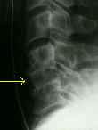
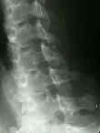
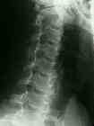
- See: Pillar View
- Discussion:
- demonstrates primarily neural foramina, pedicles, articular masses, apophyseal joints, & relative relationship at lamina;
- oblique views show the pedicle in profile, and also allows assesment of the intervertebral foramina (and osteophytes encroaching
along their margins);
- Technique:
- routine oblique views require rotating the patient's head and body;
- may be obtained in AP or PA projections:
- erect position is more comfortable;
- entire position is rotated 45 deg to one side to avoid rotational differences among different vertebral segments;
- central beam directed to C4 vertebra with 15-20 deg cephalic tilt;
- Trauma Oblique:
- this view shows the pedicles and articualr processes well;
- oblique views often are superior to any other view, including MRI or CT scans, for visualizing articular process frx & subluxations;
- in difficult question of facet subluxation, flexed oblique views also can be obtained;
- uncinate processes, pedicles, laminae, & inferior & superior articular facets are well seen using this technique;
- C7-T1 relationship, which is frequently obscured on lateral film, may be seen on the oblique and obviate need for a swimmer's view;
- Technique Trauma Oblique:
- trauma oblique series can be obtained without moving the head but by angling the tube 30-40 deg from the horizontal;
- trauma oblique is obtained w/ x-ray beam 45 deg off vertical, patient supine, & ungridded cassette horizontal & located
towards opposite side of the patient;
- this view shows pedicles & articular process well, although appearance of spine is slightly spread out;
- major benefit of oblique view is that patient can remain supine;
- no rotation of the torso or head is required;
- furthermore, oblique views often are superior to any other technique, including CT or MRI scans, for visualizing articular process
fractures and subluxations;
- in difficult questions of facet subluxations, flexed oblique views also can be obtained


