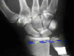
- Discussion:
- indicated for arthritic RU Joint w/ limitation of motion;
- may be useful for rheumatoid patients w/ ulnar translocation, as well as the caput ulna syndrome;
- procedure involves resection of a portion of distal ulna shaft and fusion of the ulnar head to the radius;
- maintains function of triangular fibrocaritlage complex, & maintains normal anatomic configuration of wrist
- in addition, the ECU tendon is maintained in a relatively normal position in relation to the carpus;
- procedure should restore forearm rotation;
- it should not be performed when the ulnar variance is positive ulness ulna is shortened as part of the procedure;
- in the report by Carter PB and Stuart PR (2000), the authors conducted a retrospective series of 41 Sauve-Kapandji
procedures carried out for complications of fractures of the distal radius;
- indications for surgery were pain on the ulnar side of the wrist and decreased rotation of the forearm;
- pain was improved in 25 of the 37 patients, and unchanged in ten;
- rotation of the forearm returned to within 7° of the uninjured side;
- The Sauve-Kapandji procedure for post-traumatic disorders of the distal radio-ulnar joint
- Technique:
- incision is made between the ECU and EDQ, beginning 5 cm above the ulnar head and ending distal to the ulnar head;
- care is taken to avoid cutting the dorsal ulnar sensory nerve;
- care is taken to avoid disturbing the ECU tendon sheath, and instead the dissection proceeds thru the tendon sheath of the EDQ;
- ulnar neck and proximal aspect of ulnar head are exposed;
- a towel clip is applied to the ulnar head (or a K wire is driven into the ulnar head) to help with orientation and manipulation of the distal fragment;
- osteotomy: performed just proximal to the RU joint articular cartilage, or just proximal to the flare of the ulnar head;
- a second cut is made 15 mm proximal to the first cut and the segment of ulna is removed;
- RU joint articular cartilage is removed;
- ulnar head is applied to the radius and is held with pins or w/ a 4.0 cancellous screw and a pin;
- mobilize the pronator quadratus into the defect left by the resected ulnar shaft to prevent bone bridging;
- pronator is achored by drill holes made in the dorsal side of the ulnar stump;
- dorsal capsule should be repaired;
- Complications:
- distal ulnar instability:
- if excessive bone is resected, distal portion of proximal ulna may be unstable;
- this complication is more likely to occur if instability existed preoperatively;
- a non painful clunk will be present in more than 50% of patients;
- reactive bone formation:
- if inadequate bone is resected, reactive bone may form at osteotomy site, limiting motion;
- radio-ulnar impingement
The Sauve-Kapandji procedure.
Sauve-Kapandji procedure for disorders of the distal radioulnar joint: a simplified technique.
The Sauve-Kapandji procedure for reconstruction of the rheumatoid distal radioulnar joint.
- indicated for arthritic RU Joint w/ limitation of motion;

- may be useful for rheumatoid patients w/ ulnar translocation, as well as the caput ulna syndrome;
- procedure involves resection of a portion of distal ulna shaft and fusion of the ulnar head to the radius;
- maintains function of triangular fibrocaritlage complex, & maintains normal anatomic configuration of wrist
- in addition, the ECU tendon is maintained in a relatively normal position in relation to the carpus;
- procedure should restore forearm rotation;
- it should not be performed when the ulnar variance is positive ulness ulna is shortened as part of the procedure;
- in the report by Carter PB and Stuart PR (2000), the authors conducted a retrospective series of 41 Sauve-Kapandji
procedures carried out for complications of fractures of the distal radius;
- indications for surgery were pain on the ulnar side of the wrist and decreased rotation of the forearm;
- pain was improved in 25 of the 37 patients, and unchanged in ten;
- rotation of the forearm returned to within 7° of the uninjured side;
- The Sauve-Kapandji procedure for post-traumatic disorders of the distal radio-ulnar joint
- Technique:
- incision is made between the ECU and EDQ, beginning 5 cm above the ulnar head and ending distal to the ulnar head;
- care is taken to avoid cutting the dorsal ulnar sensory nerve;
- care is taken to avoid disturbing the ECU tendon sheath, and instead the dissection proceeds thru the tendon sheath of the EDQ;
- ulnar neck and proximal aspect of ulnar head are exposed;
- a towel clip is applied to the ulnar head (or a K wire is driven into the ulnar head) to help with orientation and manipulation of the distal fragment;
- osteotomy: performed just proximal to the RU joint articular cartilage, or just proximal to the flare of the ulnar head;
- a second cut is made 15 mm proximal to the first cut and the segment of ulna is removed;
- RU joint articular cartilage is removed;
- ulnar head is applied to the radius and is held with pins or w/ a 4.0 cancellous screw and a pin;
- mobilize the pronator quadratus into the defect left by the resected ulnar shaft to prevent bone bridging;
- pronator is achored by drill holes made in the dorsal side of the ulnar stump;
- dorsal capsule should be repaired;
- Contra-indications:
- unstable RU joint or RU joint dislocation;
- Complications:
- distal ulnar instability:
- if excessive bone is resected, distal portion of proximal ulna may be unstable;
- this complication is more likely to occur if instability existed preoperatively;
- a non painful clunk will be present in more than 50% of patients;
- reactive bone formation:
- if inadequate bone is resected, reactive bone may form at osteotomy site, limiting motion;
- radio-ulnar impingement
The Sauve-Kapandji procedure.
Sauve-Kapandji procedure for disorders of the distal radioulnar joint: a simplified technique.
The Sauve-Kapandji procedure for reconstruction of the rheumatoid distal radioulnar joint.

