

-The surgical treatment of isolated mason type 2 fractures of the radial head in adults: comparison between radial head resection and open reduction and internal fixation.
- posterolateral approach: (Kocher Approach)
- approach the fascial plane between the ECU and anconeus muscle
- direct lateral approach is preferred by some surgeons because it spares the lateral ulnohumeral ligament;
- it is essential that LCL is not damaged, and hence the dissection procedes anterior to lateral ligament and anterior to the head and neck;
- at the time of surgery, look for injury to the lateral ligamentous complex, and consider operative repair if instability is an issue;
- anterior surface of the lateral epicondyle is exposed, and subsequent dissection achieves full visualization of the articular surface;
- further visualization is achieved w/ forearm pronation;
- reduction and temporary fixation is obtained w/ K wires or tenaculum clamp;
- often these frx require insertion of 2 or more screws parallel to joint line, w/ one screw placed in anterior half and one placed in posterior half;
- application of a plate requires more distal exposure;
- if stable anatomic reduction cannot be obtained, then frx fragment is excised;
- enhanced exposure:
- can be enlarged w/ osteotomy of lateral epicondyle & its reflection anteriorly with the extensor muscle origins;
- radial nerve is identified in the substance of the supinator;
- temporary fixation with K wires following reduction;
- suture anchors can be attached to the lateral epicondyle;
- Implants: (safe zone for implant insertion)
- proximal radial plate:
- dorsal distal radius plate (may be contoured to fit the radial head and neck);
- minicondylar plate;
- indicated if the head requires attachment to the neck;
- consider use of 2.0 or 2.7 mm L-shaped plate;
- screws:
- note that most radial head frx occur thru non articulating portion of radial head, which allows screws to be placed w/o having to counter-sink screw head;
- AO screws: 1.5, 2.0 or 2.7 mm cortex, depending on size of fragment;
- most often 2.7 mm miniscrews are chosen and are countersunk to avoid screw prominence;
- if a coutnersink is to be used, be sure that the screw is not too long, so as the screw tip does not extend beyond the cortical surface;
- over drilling of the proximal fragment is not required (can result in fragment comminution), especially if the fragment is held in compression during screw insertion;
- for lag screw effect, 2.7 mm drill-gliding hole is made thru the near cortical fragment, which is followed by 2 mm drill hole;
- Complications:
- posterolateral instability
- avascular necrosis:
- may occur following ORIF of comminuted fractures when most of the soft tissue attachements have been stripped;
- when this complication occurs and is symptomatic, a delayed excision may be performed once the capsulo-ligamentous structures have healed;
- non union:
- in the presentation by Ring D, et al (15th Annual Meeting of Orthopaedic Trauma Association, 1999), the authors noted 7 patients with radial head non union following ORIF on 70 patients;
- the authors point out that the limited blood supply may lead to the relatively high occurance of non union;
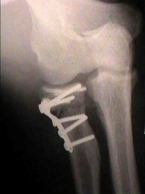
Case Example:
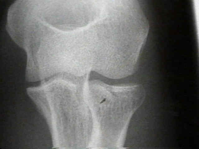
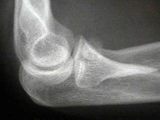
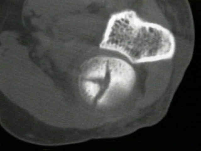

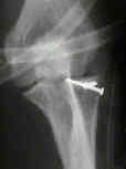
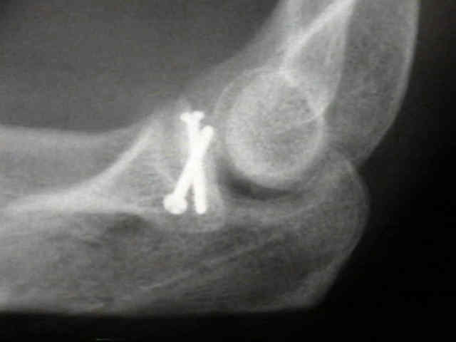
Comminuted Fractures of the Radial Head. Comparison of Resection and Internal Fixation.