

- See: Posterior Cruciate Ligament:
- Discussion:
- unlike the ACL, the PCL does have a capacity to heal;
- references:
- Chronically injured posterior cruciate ligament: Magnetic resonance imaging.
- Magnetic resonance imaging of posterior cruciate ligament injuries: assessment of healing.
- The natural history of acute, isolated, nonoperatively treated posterior cruciate ligament injuries. A prospective study.
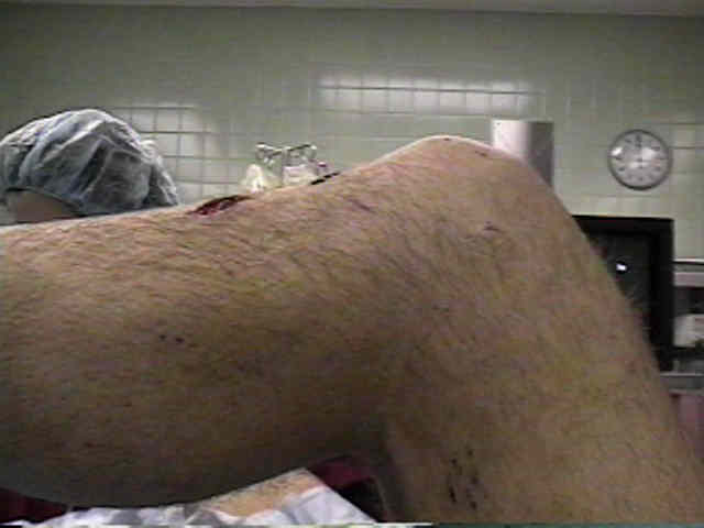
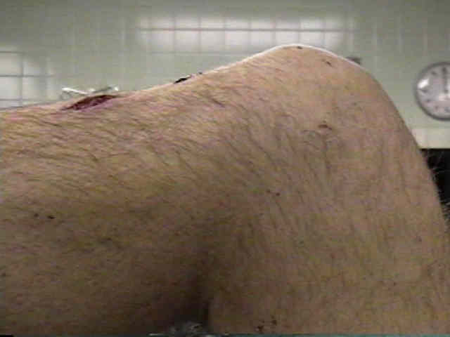
- Skin Changes:
- since PCL tears often arise from dash board injuries, there will often be contussions and lacerations over the proximal tibia;
- Quadriceps Acitve Test for PCL Injury;
- quads active test is performed by flexing pt's knee to 90 degrees, while stabilizing the foot and thigh;
- patient contracts the quadriceps while the examiner evaluates the knee for anterior tibial displacement, which is indicative of PCL;
- reference:
Use of the quadriceps active test to diagnose posterior cruciate-ligament disruption and measure posterior laxity of the knee.
- Posterior Sag Sign:
- note the amount of posterior sag relative to the other leg;
- the anterior tibial surface normally lies 1 cm anterior to the femoral surface;
- this is checked in both extension and 90 deg of flexion, and is checked in both
internal and external rotation;
- w/ posterolateral knee instability there may be more posterior sag in
external rotation;
- Posterior Drawer Test:
- probably the most accurate method of diagnosing PCL injury;
- posterior drawer test should be most sensitive for the detection of injury of PCL
when it is performed at 90 degrees of knee flexion;
- positive anterior drawer sign that is changed very little by external & internal rotation
of tibia suggests that the PCL is torn;
- w/ an isolated injury, test becomes negative as tibia is internally rotated because in this position PCL becomes taunt;
- isolated injury of PCL results in posterior translation that is increased at all angles of flexion but is max at 70-90 degrees;
- in the study by Bergfeld JA, et al. (2001), the authors evaluated the effects of internal and external tibial rotation on posterior
laxity with the knee held in varying degrees of flexion after the PCL and meniscofemoral ligaments had been cut;
- 20 knees were mounted in a fixture with six degrees of freedom, and anterior and posterior forces of 150 N were applied;
- at 30°, 60°, and 90° of flexion, there was a significant increase in posterior laxity following transection of the posterior cruciate and
meniscofemoral ligaments.
- at 60° and 90° of flexion, there was significantly less posterior laxity when the tibia was held in internal compared with external
rotation;
- at 0° and 30° of flexion, there was no significant difference in posterior laxity when the tibia was held in internal compared with
external rotation;
- internal tibial rotation resulted in significantly less laxity than external tibial rotation did at 60° and 90° of knee flexion;
- an isolated injury of the PCL is best detected when a posterior drawer test is performed with the knee in 90° of flexion;
- repeating this test with the tibia internally rotated will result in a substantial decrease in the amount of posterior laxity at 60° and 90°
of knee flexion;
- ref: The Effects of Tibial Rotation on Posterior Translation in Knees in Which the Posterior Cruciate Ligament Has Been Cut
- isolated injury of PCL would not result in change in primary varus rotation or primary external rotation;
- however, w/ concomitant posterolateral knee instability, there should be a significant increase in posterior translation, varus
rotation, and external rotation;
- tibial station:
- normal: at 90 deg flexion, the tibial should lie 1 cm anterior to the femoral condyles;
- grade I: using the normal side for comparison, a 0.5 cm difference in relative posterior translation;
- grade II: the anterior surface of the tibia and femoral condyles are flush (greater than 1 cm of relative posterior translation);
- grade III: tibia can be translated 1 cm posterior to the anterior femoral condyles;
- note that grade III instability is most often associated w/ posterolateral instability;
- references: Posterior tibial subluxation of the posterior cruciate-deficient knee.
- Posterolateral Instability:
- reversed pivot shift:
- external rotation recurvatum test detects abnormal external rotation of tibia on femur assoc w/ or in combination w/ excessive recurvatum;
- test w/ patient supine and compare it with the normal knee (simultaneously);
- move the knee from about 10 deg of flexion to maximum extension while observing and palpating external rotation of proximal
end of tibia as well as the amount of recurvatum;
- w/ complete posterolateral disruption (and intact PCL), is expected to show excessive external rotation, posterior translation, and
varus deformity, which is maximal at 30 deg of flexion;
- when external rotation is maximal at 90 deg of flexion, it is likely that there is a concomitant PCL tear;
- markedly positive test in both 30 and 90 deg of flexion indicates that PCL, posterolateral corner, and fibular collateral ligament are torn
Acute posterolateral rotatory instability of the knee.
The absent posterior drawer test in some acute posterior cruciate ligament tears of the knee.
Chronic posterolateral rotatory instability of the knee.
The accuracy of the clinical examination in the setting of posterior cruciate ligament injuries.