

- Discussion:
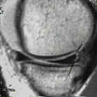
- posterior horn of the medial meniscus is difficult structure to view w/ arthroscope, esp in very tight joints with little valgus laxity;
- Positioning: Valgus Stress to the Slightly Flexed Knee:
- valgus stress applied to slightly flexed knee (no > 10 deg) combined w/ external rotation of tibia allows visualization of posterior horn of the medial meniscus;
- when valgus stress is applied to slightly flexed knee while tibia is externally rotated, direct manual pressure can be applied at level of posterior joint line to push meniscus anteriorly;
- mistake is to attempt to open up medial joint w/ too much flexion;
- Technique:
- for access to posteior third of medial meniscus, the instrument portal should be placed just anterior to the medial collateral ligament at level immediately above the superior surface of the mensicus;
- more resistant the joint is to medial opening, the more important it is to place the instrument portal precisely anterior to MCL;
- w/ more anterior portal site, medial femoral condyle will obstruct access to the posterior meniscal segment;
- if moderate valgus laxity is present, posterior segment of meniscus can be reached thru an anteromedial supramensical portal;
- farther the medial joint space can be stressed open, the more anteriorly the instrument portal may be placed;
- in tight joints, two instrument portals are often necessary for adequate treamtent of the meniscus
Arthroscopic posteromedial visualization of the knee.