

 - See:
- Anastomoses of Lower Limb Arteries
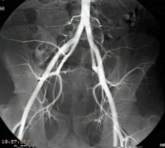
- Femoral Arteriogram:
- Lower Limb: anatomy of arteries and bones with 3D pictures and angiographic radiological images
- Anatomy:
- common iliac bifurcates at the L5-S1 disc;
- internal iliac:
- courses anterior and adjacent to the sacroiliac joint;
- posterior branches of the internal iliac artery: iliolumbar, superior gluteal, and lateral sacral arteries;
- anterior branches of internal iliac artery: obturator, umbilical, vesical, pudendal, inferior gluteal, rectal, and hemorrhoidal arteries
- external iliac artery passes obliquely down medial border of psoas & anterior and lateral to external iliac vein;
- external iliac artery becomes the common femoral artery as it passes below the inguinal ligament;
- common femoral artery is approximately 4 cm in length and divides into superficial femoral & profunda femoris arteries;
- in upper thigh, this artery lies between femoral vein & nerve in femoral triangle, space roofed by fascia lata
and bounded by inguinal ligament above, satorius muscle laterally, & adductor longus medially;
- largest branch of the femoral artery in femoral triangle is profunda femoris, which arises on lateral side of the femoral artery, arches posteriorly,
and continues downward near the middle of the thigh;
- descending genicular artery arises from femoral artery;
- at the distal apex of femoral triangle, above the knee, it passes thru opening in adductor magnus to enter popliteal space as popliteal artery;
- after providing genicular arteries at level of knee joint, it passes deep to soleus, where it transverses thru another fibrous tunnel; (hence, artery remains
vulnerable during dislocation of knee because of tethering);
- popliteal artery then sends paired sural arteries to gastrocnemius & soleus & ends by dividing into anterior & posterior tibial arteries;
- Femoral Artery Thromboembolectomy:
- longitudinal groin incision;
- common, superficial, and deep femoral arteries dissected free and controlled with elastic tapes;
- transverse arteriotomy just above the femoral bifurcation;
- extraction of thromboembolus from SFA with a 4 French Fogarty balloon
- deep femoral artery clot removal follows with a 3 or 4 French Fogarty
- back bleeding from the SFA controlled by traction on elastic tape or by a traumatic vascular clamp
Femoral artery thrombosis after open reduction of an acetabular fracture.
Acute trauma of the femoral artery and vein.
Superiority of the femoral artery of monitoring. A prospective study.
Prospective evaluation of radial and femoral artery catheterization sites in critically ill adults.