

- Discussion:
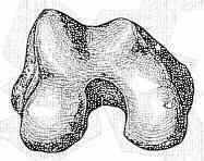
- when viewed in cross section, shape of distal femur resembles trapezoid w/ medial side inclinded about 25 deg & lateral side about 10 deg;
- both the seating chisel and the subsequent blade plate should be 1 cm to 2 cm short of medial cortex to prevent inadvertent penetration;
- this is due to the trapezoidal shape of the femoral condyles, which are narrower anteriorly than posteriorly;
- posterior diameter is longer than the anterior;
- therefore a plate which appears to be just the right length on AP view will be too long & will penetrate cortex and
protrude deep to the MCL or become subcutaneous;
- correct length is 15 to 20 mm less;
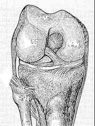
- anterior surface slopes downwards to medial side and corresponds in inclination to the patellofemoral joint;
- care must be taken therefore that any device inserted is parallel to this inclination or it will end up in patellofemoral joint;
- when distal femur is seen on lateral view, it is seen that femoral condyles appear to have been added on posteriorly to the shaft;
- therefore, target for plate insertion is in middle of anterior half of condyles